Feeds:
Posts
Comments

One of the most recent advances in cancer therapy has been the development of new drugs inhibiting a class of enzymes called tyrosine kinases.  Normally, these enzymes are involved with the transduction of signals involving the growth and proliferation of a cell.  In cancer cells, however, their activity is greatly increased such that they gain increased ability to proliferate and spread general nastiness, in contrast to the ordered and regulated growth of normal cells.  In 2005, one of the first drugs of the class, imatinib, was lauded by time magazine and the New York Times as a great leap forward in cancer treatment and “converted a fatal cancer into a manageable chronic condition.” [NYtimes]  With few treatment options besides a bone marrow transplant, chronic myelogenous leukemia was one of the more fatal kinds of cancer.  However, with these new therapies researchers have shown that “after 60 months of Gleevec (imatinib’s brand name) therapy, 98% of patients had shown a complete hematologic response. Also at 60 months, the estimated overall survival rate for patients was 89%, with a relapse rate of only about 17%.” [1]

However, a word of caution must be spread.  These drugs are not the often sought after ‘cure for cancer,’ as patients still run the risk of relapse and mutation fueled resistance to the therapies, as well as the controversy considering the incredible cost of treatment (imatinib costs $32-98K per year for CML).  That said, their development is still a milestone in our understanding of cancer pathology and its treatment.  Much of the class was developed out of what is called ‘rational design’ methodology, that is to say by using deductive means based upon knowledge of the shape and function of the principal components of the systems.  This is opposed to drug screening strategies, used during much of the earlier history of drug discovery, of finding a lucky diamond in the rough and modifying it to fit certain ‘structural activity relationships’.  Direct knowledge of the target areas and their effects has allowed medicinal chemists to design new approaches from the ground up to develop more effective treatments for disease.  Perhaps, as the knowledge base becomes deeper and more comprehensive, we might be able to attack the real root of the issue in this terrible disease, but for the time being we must be patient with the fruits that our limited knowledge provides.

What exactly is a tyrosine kinases and what does it do?

Tyrosine kinases are enzymes that act as chauffeurs of a chemical signal, that when passed along to other mediaries can effect some kind of action in a cell.  These actions are many and varied.  In relation to cancer, these are typically signals to allow growth, cell division or the creation of signals to induce the production of new blood vessels.  When activated, tyrosine kinases usually pass the signal down to relay proteins that activate the downstream machinery for these actions, in effect acting as an ‘on/off switch’ for that particular activity.

Cells have many different kinds of RTKs and we must distinguish between two different general classes when considering these drugs.   Receptor tyrosine kinases are membrane bound receptors for a long list of growth factors, that in response to the extracellular binding of the factor, the receptor becomes activated and allows the association with other signaling proteins by self-phosphorylating tyrosine residues in the trans-membrane and C-terminal regions of the protein’s structure.  These create docking spaces for downstream signaling proteins, like Ras, STAT and PI3K, allowing larger signaling complexes to form and the information to be passed on.  Much like a Dremel tool with a selection of interchangeable parts, these kinds of signaling proteins can modulate the signal sent and create a whole network of interactions within the system.  For example, when a Ras protein is activated by the receptor interaction can induce a MAP kinase cascade that culminates with protein transcription in the nucleus by unlocking a large set of transcription factors.  PI3K is involved with cell survival and insulin induced metabolic control.  Likewise, STAT proteins can be activated in this fashion and move into the nucleus where they become active transcription factors.  There are also non-Receptor tyrosine kinases, which are usually located in the cytoplasm or nucleus instead of the cell membrane.  Two of the most common are c-src and abl, which are involved in a similar array of growth regulating, cell maintaining activities.  The abl tyrosine kinase, for example, is usually located in the nucleus and is involved in the repair of DNA damage and regulation of apoptosis and transcription.  Src is involved heavily in the regulation of cell division and differentiation in many cells.

Where cancer comes in is the fact that some mutated or viral versions of these enzymes are constitutively active, that is to say, stuck in the on position.  This activity is in part responsible for the explosive growth seen in some tumors.  Some cancer inducing retroviruses, like the Rous sarcoma virus or murine Abelson leukemia virus, are known to produce these kinds of kinases as part of their payload.  In fact, the first tyrosine kinase to be discovered was called ‘v-src kinase’, denoting where it had come from.   The WHO has estimated that around 17% of all human cancers might be caused by these kinds of rouge viral kinases. [2] Furthermore, in 1972 Janet Rowley noticed that patients with a rare and deadly form of cancer called chronic myelogenous leukemia had a chromosomal abnormality later named the Philadelphia chromosome.  This was caused by the transplantation of two genes ‘abelson tyrosine kinase’ and ‘break point cluster’ creating a chimeric gene of the two called Bcr-Abl.  The fusion protein created by this gene is another ‘always on’ version of the enzyme and was where these new therapies got their start.  Though these enzymes are ubiquitous and normal cells do have an Abl kinase, Bcr-Abl is unique enough that drugs can be targeted towards it and other cell kinase redundancies can cover for its loss of function.  This affords a more specific therapy against these cancer cells than other earlier chemotherapy based strategies.

Where da Drugs At?

Since the tyrosine kinases require ATP for activation, quite a few of these compounds were developed from the idea to ‘poison the well’ with a similar looking, yet difficult to remove compound at the ATP binding site.  The early research drug semaxanib was developed for this purpose and led to the development of the ‘non-receptor tyrosine kinase inhibitor’ sunitinib, and the first ever dog specific anti-cancer drug: toceranib (Yeah, you read that right, “priorities”).  Each of these has similarity with the structure of ATP built right into the compound, which is close enough to enable them to bind to the enzyme’s active site.

Though semaxanib is a natural product, the majority of these compounds are purely synthetic and were developed through direct knowledge of the enzyme structure and its interactions.  Knowledge of the binding space has allowed designers to make judicious choices in functional groups to elicit specific bonding to the enzyme.  One interesting point of note is the use of highly electronegative groups, like fluorine atoms, to anchor and bind to amino acids in the enzyme’s superstructure.  The reasoning is twofold in that this enables better targeting of the compound to the receptor space via interaction with polar amino acid groups and makes them more difficult to remove from the enzyme in natural conditions.  The drugs regorafenib and sorafenib are good examples of this, which include a trifluoro-methane group (-CF3) that is intended to anchor these compounds in the right place.

Considering current human therapies, first line therapies inhibiting cancers involving mutations to epidermal growth factor receptors, like erlotinib and gefitinib, have long been newsworthy.  Therapies attacking non-receptor tyrosine kinases, like abl kinase and it’s CML fusion cousin, are exemplified by Imatinib, dasatinib and nilotinib.  These are targeted to block excessive receptor signaling by damaged kinases of these receptors.  .  Resistances, however, are apparent even with these new therapies, as a single point mutation in the gene encoding the enzyme removes their effectiveness and reinstates bcr-abl activity.  The T315I point mutation changes a single amino acid (threonine to isoleucine) in the enzyme and is the most prevalent resistance found among resurgent CML, occurring in perhaps “6 out of every 9 cases of imatinib resistance.” [3] This mutation confers resistance to not only imatinib, but also others of the class, due to its critical location guarding the binding pocket of the enzyme.  Dasatinib and nilotinib represent the second generation therapies over imatinib, which, in the case of nilotinib has 30 times the binding efficacy in imatinib resistant cancer cells at the bcr-abl binding site.  Furthermore, in the case of non-small cell lung carcinoma, a small percentage of people have a similar mutation creating a fusion protein called EML4-ALK, which is similarly specifically targeted by the drug crizotinib. [4]

Vascular endothelial growth factor (VEGF) is involved with the new production of blood vessels around a tumor that provide if with the additional blood flow and nutrition needed for increased growth.  This “angiogenesis” has long been implicated as a critical event in the development of deadly tumors and prognosis can even be intuitively estimated by the amount of VEGF in a sample and thus vascularization of a given tumor.  As luck would have it, VEGF receptors have in common a tyrosine kinase that we have recently learned to inhibit with the drugs pazopanib, axitanib and vandetanib.  Pazopanib has been approved for use against renal cell carcinoma and has shown moderately positive results in phase II studies involving its use against soft tissue sarcomas and ovarian cancer. [5] Axitanib has been also tested against metastatic renal cell carcinoma with positive results as: “axitinib increased median progression-free survival time by 43% among the overall patient population, from 4.7 months to 6.7 months (maximum 8.6 months). Importantly, benefits of the drug were evident in patients who had previously been treated with cytokines or sunitinib.” [6] Vandetanib has likewise been tested against a kind of thyroid cancer with the “estimated progression-free survival for the patients treated with vandetanib was 30.5 months.” [7]

Final thoughts

Though our knowledge of cancer pathology has clearly increased to point to effectively manipulate specific areas of weakness in the cancer cell machinery, developing resistances and the sheer cost continue to remain a challenges for widespread effective therapy.

[NYTimes] Interesting NY Times interview with Druker.

[1] “After 5 years of follow-up, continuous treatment of chronic-phase CML with imatinib as initial therapy was found to induce durable responses in a high proportion of patients.”

[2]The estimated total of infection-attributable cancer in the year 2002 is 1.9 million cases, or 17.8% of the global cancer burden. The principal agents are the bacterium Helicobacter pylori (5.5% of all cancer), the human papilloma viruses (5.2%), the hepatitis B and C viruses (4.9%), Epstein-Barr virus (1%), human immunodeficiency virus (HIV) together with the human herpes virus 8 (0.9%). Relatively less important causes of cancer are the schistosomes (0.1%), human T-cell lymphotropic virus type I (0.03%) and the liver flukes (0.02%).”

 [3] CML point mutations study.

 [4] “The primary endpoint of both trials was objective response rate (ORR) as assessed by the investigator. In Study A, the ORR was 50 percent (95 percent CI: 42 percent, 59 percent) with a median response duration of 42 weeks. In Study B, the ORR was 61 percent (95 percent CI: 52 percent, 70 percent) with a median response duration of 48 weeks. Complete responses were observed in 1 percent of patients. No differences in ORR by performance status, number of prior chemotherapeutic regimens, or percentage of cells found to have the ALK gene rearrangement were noted.”

[5]   Admittedly, this is a shitty source without links to useful information, but hey, at least it has impressive looking numbers that look like it has information to back up its claims.  I’m very sorry for including this in here, as laziness has gotten the better of me.

[6] “The Phase III Axis 1032 study included 723 patients, who were randomized to receive either axitinib or sorafenib twice daily.  54% of patients had previously been treated using sunitinib-containing regimens, and 35% had received prior cytokine-based therapy. The results showed that compared with sorafenib therapy, axitinib increased median progression-free survival time by 43% among the overall patient population, from 4.7 months to 6.7 months (maximum 8.6 months). Importantly, benefits of the drug were evident in patients who had previously been treated with cytokines or sunitinib, Pfizer notes.

In previously cytokine-treated patients, axitinib therapy led to a median progression-free survival of 12.1 months, compared with 6.5 months for the sorafenib group. Among previously sunitinib-treated patients, progression-free survival was 4.8 months for those moved on to axitinib therapy, compared with 3.4 months for those given sorafenib. Secondary trial endpoints showed that the independently assessed objective response rate was 19.4% among the overall axitinib patient cohort, compared with 9.4% for the sorafenib cohort. “

[7] (http://www.cancer.gov/clinicaltrials/results/summary/2011/vandetanib1211)

“It is demonstrable that things cannot be otherwise as they are; for as all things have been created for some end, they must necessarily be created for the best end.” –Pangloss

Tragedy struck on December 27, 2008 when a prominent AIDS denialist succumbed to complications from her disease. Christine Maggiore had a long and successful career denying the HIV virus causes AIDS, with a high point surely her advisory role in South Africa’s temporary block on government funded AIDS medical treatment (estimated to have cost the lives of 330,000 people), or perhaps the founding of “Alive and Well AIDS Alternatives,” an organization dedicated to encouraging HIV infected people to avoid HIV medications and treatment in favor of naturopathic remedies. I don’t personally believe she was a good person, but one can’t deny she suffered because of her beliefs and the choices she made considering them. To be equally fair one must also consider how they affected the lives of the people closest to her.

Great controversy was created with the publicizing of her choice to breast feed her children despite being tested HIV positive. Her public stance to not take antiretroviral medication while doing so increased the likelihood of transmission to her daughter. These drugs have been shown to reduce this risk, with the drug AZT thought to reduce it by at least 23% [1]. At the age of three, her daughter Eliza Jane fell ill with Pneumonia-like symptoms and her mother’s continued refusal to have her tested for HIV made matters worse. After doctor shopping, she eventually chose a sympathetic holistic practitioner connected with her organization for treatment who claimed her daughter was “only mildly ill.” A month later she was dead. Autopsy reports revealed a more disturbing picture showing that she was seriously underweight, had pronounced atrophy of the thymus and lymphatic organs and her lungs were infected with an opportunistic pathogen called Pneumocystis jiroveci (#1 cause of death in pediatric AIDS cases), all of which are considered classic indicators of AIDS. Furthermore, it revealed HIV protein components in her brain tissue, which is an indicator of HIV encephalitis. Maggiore rejected the conclusions alleging fraud and incompetence, choosing instead to rely of the review of an animal pathologist close to her organization. Predictably, his findings described her death as attributed to a poor interaction from amoxicillin, a claim dismissed as preposterous by professionals in the field. Ultimately, Maggiore would not have long to consider the nature of her daughter’s death, as her own health began to take a severe turn for the worse.

I don’t believe there is anything funny or ironic in this tragedy, just sadness and pity. A three year old is dead and died needlessly, infected as a direct result of her mother’s inconsiderate beliefs. Thousands more died as a result of her enterprise and even more are continually mislead into unsafe and disreputable treatments, hampering real efforts to contain and manage the disease. What went so wrong, that would force her to ignore all medical evidence regarding her condition? What made the collective body of evidence so thoroughly unconvincing, so unbelievable that she would gamble the lives of her children upon this rhetoric? The inability to convince the majority of people or to provide a successful, publicly accepted counter-argument to this surely is a great collective failure of medicine today. Despite its successes treating injury and establishing a biological basis for disease there are many that whether of stubborn obstinance or more likely lacking a clear description of the science behind it find it inconsistent or at worst disreputable.

Anti-vaccinationists, for example, despite years of successful treatment and the virtual eradication of diseases like small pox and polio

Old cows still say moo.

in the western world, still pose arguments of the dangers and inefficacy of the treatment. Religious and personal freedom based arguments posed reach back well over 100 years to echo the originals. However, arguments alleging the dangers of it are finding fresh audiences despite both empirical and otherwise anecdotal evidence to the contrary. Peter Morrell, a UK homeopath, wrote in a scathing editorial in 2000[2]:

“It does not work; it is unnatural, that the human race has survived healthily for countless generations without them and that homeopathy provides a better alternative that is both safer and effective…One can see the dangers and pitfalls of vaccination as another Russian roulette game not worth the risk.”

This is probably safe.

Perhaps if the collective medical profession relied upon a farcical theory like homeopathy’s “water memory,” then the comparison to gambling would be apt, but this is not the case. Though it is impossible to account for every possibility, medications and treatments are tested for both safety and effectiveness. Furthermore, results are verified by repeated testing to further clarify specific effect from lucky incidence. The double blind trial developed in the early 1900’s further limited sources of error such as observer bias and experimenter effects. It did more to help the sick in 100 years, than ostensibly “better alternatives” did in 200, by allowing the measurement of an effect to be defined and separated from statistical placebo effects. If homeopathy truly provides a better alternative, then results from studies [3][4] would clearly show benefit beyond placebo and experiments with positive correlation would be repeatable by other parties. After all, what good is a treatment that works only for some or some of the time for a few lucky people? If we’re seriously going to make the allusion to gambling, wouldn’t it be better to side with the hand the yields a consistent and high rate of return?

“Infectious diseases in general have declined massively since 1900…”

It also shouldn’t surprise anyone who has taken a bath recently, that good hygiene is good preventative medicine. Before the advent of sewage systems, cities were severely limited in size, usually by the occurrence of disease. John Snow’s 1854 correlation that the year’s cholera epidemic was caused by contaminated city water (specifically around a single city pump) established a basis for statistical observation in the causes of disease. Though he did not know the exact cause of the cholera, using a map he was able to show that nearly all of the deaths attributed to the epidemic could be traced back to that particular location. It was later shown that the pump was dug too close to a cesspool and it was leaking fecal matter into the drinking water. Cleanliness and proper hygiene have an effect on certain diseases, yet it is important to make a distinction that it does not affect all diseases in the same fashion. This can be seen even today with outbreaks of measles and rubella (etc) among unvaccinated populations in western nations despite the herd immunities largely present and public water purification efforts taken. The MMR vaccine controversy in the 2000’s started with the speculation that the vaccine could cause autism and continues to cause great fears among parents pushing a dip in non-compliance. This dip has been credited with causing a large upsurge in these diseases. The CDC, for example, reported that cases of measles in 2008 were at a 12 year high [5][6]. Publicly, it matters little that the progenitor of this vaccine scare, Dr Andrew Wakefield, has been shown to have altered his data in favor of parties he accepted money from [7][8]. To the public, the fear is real and the accusation is enough to cause panic.

Royal Rife hard at work.

I recently had the pleasure of interviewing a UFO conspiracy theorist. His main argument was that all sciences, except aeronautics, have advanced exponentially[*] since the Roswell UFO landings, perpetuating unearthly advances in science and technology that were previously impossible with our ‘feeble’ intellect. There is a fallacy here that doesn’t revolve around the usual skeptics rallying point, but that there is a strong public belief in the infallibility of science, such that lay people often times, when confronted with the limitations of science, would rather believe in a conspiracy of failures than in the truth: that at any given point science is far from solving the collective mass of problems affecting us at present (“for every problem solved there are hundreds more questions”).  His argument views the discoveries of science as a linear progression, completely ignoring the ‘fallen warriors’ left by the roadside to get to that point.  The theories of phlogiston and geocentrism no longer hold the same philosophic sway they once did, as they have been replaced by more accurate descriptions of the world around us.  Much in the same way, medical science in some ways has been the victim of its own success, in that people seemingly have a genuine expectation that “a disease with a name will have a cure.” This leads to disappointment at its failures and current limited ability to cure and is exemplified by the many conspiracy theories involving “big pharma” holding back “real” treatments for AIDS and cancer. Take for example, author Barry Lynes’ 1987 book “The Cancer Cure that Worked,” about Royal Rife’s supposed cancer curing radionic devices and their subsequent cover-up [8]. Robert Strecker’s work on the alleged manufacture of the HIV at Ft. Dietrick as a tool to limit the industrial growth of Africa, as well as several involving the chemical AL-721 as an AIDs cure too cheap to sell are all examples of this common theme. The secret wish is the promise of success, in that things are as they are, because they are “created for the best end.”

The main issue is that the representatives of science have been sending a mixed message, especially in light of the recent inclusion of “alternative medicines” like homeopathy into the pantheon of medical techniques. The term itself insinuates a duality of being as if the two occupy separate halves of a holistic continuity (good/evil, yin/yang, republican/democrat). This image is only furthered by state required licensure of practitioners, yielding a further sense of legitimacy to these otherwise unproven techniques. This duality, at least in the public eye, gives a degree of plausible deniability. Science may say the evidence shows they work no better than placebos, but they can say (and do) that the scientific method is incompatible with their art. If they’re considered, even just rhetorically at an equivalent level, then it appears as just two rival businesses competing for customers and any debate on the subject is subsequently viewed as a rhetorical advertisement rather than a statement of just the facts. Legitimate studies, whether for or against, are viewed with contempt and as the product of that party’s particular bias. This issue is further compounded as medicine becomes less reliant on the humanistic bedside manner that became a doctor’s stereotypic image in the public eye. The doctor-patient relationship and the trust that comes with it has been eroded by impersonal, corporate business practice and this bubble is being largely filled by alternative medicines. People do not like being treated as a mechanistic collection of systems and holistic imagery relates to them in a way that an impersonal blood test cannot, even if the two are at odds factually. The growing impersonal aspect of modern medicine only encourages people like Maggiore and Morrell, further allowing them a gray area to exploit.
=========================================

[1] Basic and Clinical Pharmacology 10th Ed., Bertram G. Katzung, MD, PhD

[2] http://epe.lac-bac.gc.ca/100/201/300…ue-7/10-13.htm

[3] “Homeopathy remains one of the most controversial subjects in therapeutics. This article is an attempt to clarify its effectiveness based on recent systematic reviews. Electronic databases were searched for systematic reviews/meta-analysis on the subject. Seventeen articles fulfilled the inclusion/exclusion criteria. Six of them related to re-analyses of one landmark meta-analysis. Collectively they implied that the overall positive result of this meta-analysis is not supported by a critical analysis of the data. Eleven independent systematic reviews were located. Collectively they failed to provide strong evidence in favour of homeopathy. In particular, there was no condition which responds convincingly better to homeopathic treatment than to placebo or other control interventions. Similarly, there was no homeopathic remedy that was demonstrated to yield clinical effects that are convincingly different from placebo. It is concluded that the best clinical evidence for homeopathy available to date does not warrant positive recommendations for its use in clinical practice.”

[4] Another Homeopathy study.

[5] http://www.medpagetoday.com/Infectio…sDisease/10629

[6] “A trend which continues into 2009” was what I wrote, but this appears to be wrong according to the guardian. Touche~

[7] http://www.medpagetoday.com/Pediatrics/Autism/12850

[8] http://www.ncbi.nlm.nih.gov/pubmed/17867721

[*] He was an interesting character. Among other things he believed that a class of human beings were alien-human hybrids and ordinary humans lacked the intellectual fortitude to build the sphinx and pyramids, ascribing a mystical super-scientific power to their magnificence. Aeronautics was supposedly held back, because “they” want to kept us from knowing where we really come from. This is also the reason we haven’t been back to the moon, that and they have a base on the dark side. I enjoyed the interview. He even laughed when I asked if phrenology would benefit greatly from the alien wisdom. These are a different story, though.

[9] description of the book and summary.

Written on April 15, 2009

Muscular dystrophies are a class of degenerative diseases characterized by progressive muscular weakening, myopathy and paralysis.  Among these, one of the most famous is Duchenne muscular dystrophy (DMD), which affects about 1 in every 3000 males born in the United States.  This class of diseases faces the very limits of medical therapy, as there are currently no clinical treatments available to halt the progression of symptoms.  With only supportive care available, patients are left to progressively worsen, typically ending with loss of physical mobility, the ability to breathe independently and usually fatal heart related complications.  The disease maligns quality and length of life, such that most only live into their twenties and are confined to a wheelchair around their teens.  Only in very rare circumstances have any lived into their 30’s or 40’s.

It wasn’t until fairly recently that an accurate understanding of the pathology involved became clearer.  With DMD, mutations from reading frame shifts in the gene producing the dystrophin protein were found to be the root of the problem.  Dystrophin is a critical protein involved in muscle structural support and intracellular signaling with the extra-cellular matrix.  For any kind of contraction, a muscle must have an anchor and this protein helps play this part by linking the strand to a support structure called the DAP complex, which in turn links to the extracellular matrix.  Like a car without shock absorbers, muscle cells are more susceptible to physical membrane damage and slowly whither under the stress.  Damaged, leaky membranes are a classic indicator of failed cell viability and seen as the hallmark of dying cells, a fact corroborated by typical diagnostic staining procedures using cell impermeable dyes.  Dystrophin is also heavily involved with intracellular signaling from responses outside the cell.  Generally, proper signaling is necessary for continued cell viability.  Without it cells are usually marked for termination by immune system cells like macrophages, which seek out this kind of aberrant signaling.  DMD affected muscles are often infiltrated by these kinds of cells, which further increase the damage by initiation of an inflammatory response.

One of the hallmarks of this disease can be seen in slides of affected muscle.  Healthy cells are ensheathed by a thin membrane called the endomysium, but in diseased tissue the spaces around the muscle cells are filled increasingly with fat or fiber-like cartilaginous tissue.  This is called endomysial fibrosis.  As macrophages realize something is wrong, they infiltrate the area and initiate an immune response causing inflammation.  This further weakens the damaged cells and whittles the number of them down slowly, which accounts for the progressive weakening pathology of the disease.  The dead cells are typically replaced by this white tissue matrix.

Many hypothesized early on that gene therapy held the greatest promise to cure this disease.  After all, in order to truly cure a disease of genes, you must fix those broken genes.  Though pharmaceutical treatments reducing immune system response to the disease may improve conditions temporarily, they most likely won’t offer the long-term increase in survivability sought after for so long.  As such, many new methods and techniques have been proposed as models for new gene therapy based strategies.  Many of these have proven successful in mouse models and a few have even had some success in small clinical trials.  These cover a wide range of areas and strategies, such as using viral vectors for gene transplantation, up-regulation of suitable endogenous genes and forced expression of tailored mini genes.  Each of these has found success as a possible route towards a therapy, but each with its own drawbacks and limitations.  The master stroke, it seems, is still some time off, but even closer and within our child-like grasp at the cookie jar.

  1. Viral vectors and transgene infection

A now famous example comes from researchers at the University of London.  They worked with viral vectors to introduce a dystrophin replacement gene into muscular dystrophic mice.  Viral vectors are complicated, because they can provoke a response from the immune system, can possibly cause disease and have a limited storage capacity for any potential gene transplant.  They found a balance with the virus called Adeno-associated virus (AAV).  These viruses are able to infect many kinds of cells, can be produced without their endogenous viral genes, have never been shown to cause a human disease and also require a helper virus to replicate themselves after infection.  This combination of characteristics limits the dangers associated with infection to a degree and makes them good candidates for use in human gene therapies.

One of the main problems they faced was the small storage capacity the virus could carry.  The dystrophin gene is one of the largest known (2.4 megabases), so they had to make some kind of compromise.  They worked around this by manufacturing a mini-gene that was much smaller than the actual gene that coded for the dystrophin protein.  This was made possible by knowledge of the genomes of those affected by the disease.  A close cousin of Duchenne muscular dystrophy (DMD) is called Becker muscular dystrophy (BMD) and is characterized by a lesser severity of symptoms.  Surprisingly, the gene mutations in BMD, though sometimes ‘affecting ~ 50% of the gene itself’, affect much less critical locations than mutations in DMD.  Mapping these locations allowed them to whittle out less critical pieces of the DMD gene.  They developed a mini-gene that would produce a lesser functional, but still useful dystrophin protein, which was still able to fit within the size constraints of the viral carrier.

Once the mini-gene was completed they paired it with a transcription promoter from the cytomegalovirus to force gene transcription and production of the protein once the virus infected a cell.  They injected the virus into mice expressing a DMD phenotype and found that the mini-gene was successfully expressed in “greater than 50% of the muscle fibers 20 weeks after infection”(S).   It was found that it relieved aspects of DMD pathology, particularly rebuilding the DAP complex and improved muscle structure.  This culminated with mice test groups showing characteristics of a lesser severe form of the disease.  Furthermore, experiments showed that even with a low 20-30% level of gene expression, there was a substantial reduction in DMD pathology overall with the use of this mini gene.

2.       Increasing transcription of gene with artificial constructs

As it turns out the dystrophin protein is not the only one utilized to link the muscle to the extracellular network.  Before birth, another similar protein called utrophin is used preferentially for the same purpose, but is replaced almost entirely by dystrophin after birth. The protein exhibits 80% similarity with dystrophin and remains expressed during the course of the disease.  Researchers at the Istituto di Neurobiologia e Medicina Moleculare in Rome surmised that if preferential expression of utrophin was re-established it might provide a route to a cure or a reduction of symptoms to make it manageable.  Using an artificial transcriptional element called “Jazz”, they were able to restore muscle integrity and prevent the development of DMD in mice test groups.

It has long been known that the sudden expression of new, ‘foreign’ proteins runs the risk of causing an immune response as immune cells target these new proteins as antigens.  So just giving a person an extra supply of dystrophin won’t work as a treatment for the disease.  It was found that when a cell lacks proper functioning dystrophin, it up regulates utrophin to compensate, but the level is insufficient to prevent disease progression.  Since this protein is already expressed increased production could reduce complications caused by immune system interference.  Many previous studies have confirmed that: “Increased expression of utrophin restores plasma membrane integrity and rescues dystrophin-deficient muscle in mdx mice.”(S)

A ‘zinc finger’ is a recognition element that can interact with specific sections of DNA.  In this study, an artificial zinc finger was manufactured to correspond to a section of the promoter in the utrophin gene in both the mice and human genes.  Upon testing, they found that it was able to successfully bind to its specific DNA target sequence and increase production of utrophin expression to 1.8 times that of controls.   This increase in production also translated to a therapeutic benefit in mice test groups, showing increases in muscle size, fiber regeneration and lower serum levels of creatine kinase, a chemical identifier of muscle necrosis.

Possible benefit was further characterized by in vitro testing of muscles using electro stimulation.  Weak muscles, deficient of contractile force are a hallmark of DMD, yet those treated with the ZF ATF “Jazz” showed the opposite in excised diaphragm and extensor digitorum longus muscles.  These were able to perform longer and more sustained contractions, than diseased control groups.  Membrane integrity was also tested by staining with procion orange dye.  This fluorescent dye is usually only taken up into a cell with a leaky membrane, so it is often used to assess membrane integrity.  Sustained contractions to a muscle with a DMD phenotype would cause membrane damage and usually exhibit a greater dyed area than that of a healthy cell under the same conditions.  Muscles tested and stained under these conditions showed positive results for Jazz treated test sets, with greater dyed areas observed in the DMD cells.

3.       Antisense Oligionucleotides:

Antisense oligionucleotides are a class of nucleic acids that have also been tapped as a possible route to a DMD therapy.  In 2008, researchers at Oxford published a study testing the hypothesis that these could induce at least partial dystrophin protein expression by pushing the reading frame over in mutated muscle cells.  The idea in this technique is to use a technique called ‘exon skipping’ to push the ‘out of reading frame’ portions of the protein back into the reading frame and produce a partially functional, “becker-like” dystrophin protein.  This technique was shown to have benefit in not only mice, but also in humans in a proof of concept test in 2007.

Up until now, universal changes in expression from AOs were difficult to attain, with high percentage expression limited to skeletal muscle groups, but only limited expression in critical heart and diaphragm muscles.  This was a huge limitation to its possible use as a therapy, as “cardiomyopathy is a significant cause of morbidity and death in DMD patients” (S).  Without a corresponding effect upon heart muscle, any increase in overall muscle dystrophin expression would only exacerbate any heart conditions a DMD patient (or mouse) might have.  However, this new test was different and achieved much more favorable results in mice.  The question is: what did they do different?

It was surmised that the previous tests of AOs had limited entry into exclusive, protected environments like the heart.  They needed a tool that would allow access to these areas without destroying the gains made in earlier tests and this was achieved by conjugating the AOs to an arginine rich peptide scaffold.  Arginine is a positively charged amino acid and its use in the peptide yields an overall positive charge.  These kind of chemicals are thought to use special cell-mediated uptake systems in common with glycosaminoglycans, thus like a Trojan horse they facilitate the entry of whatever cargo they might bring.

Three weeks after injection, “between 25 and 100% of normal dystrophin levels had been restored in body-wide skeletal muscles” and “even in the diaphragm almost 25% of normal dystrophin protein was restored.”  Restoration was also seen in the heart, but not quite as high as that of skeletal muscle: “levels between 10 and 20% of that found in normal mouse heart were typically seen in all western analysis in all treated animals.” These results also corresponded with a function increase in muscle contractility and lower serum creatine kinase levels, both indicators of improved muscle ability and reduced muscle damage.

To build a tower on the sea.

Though these seem like giant steps towards a cure for one of the great diseases of man, there will undoubtedly be more questions than answers.  These are exciting times.

Every one of these people worked really hard. Please read these sources first hand (at least these):

1. “Adeno-associated virus vector gene transfer and sarcolemmal expression of a 144 kDa micro-dystrophin effectively restores the dystrophin-associated protein complex and inhibts myofibre degeneration in nude/mdx mice. Stewart A. Fabb, Dominic J. Wells, Patricia Serpente, george Dickson.  Human Molecular Genetics, 2002, vol. 11, No. 7, pgs 733-741.

2. “Expression of human full-length and minidystrophin in transgenic mdx mice: implications for gene therapy of Duchenne muscular dystrophy.” Wells DJ, Wells KE, Asante EA, Turner G, Sunada Y, Campbell KP, Walsh FS, Dickson G.  Hum Mol Genet. 1995 Aug;4(8):1245-50.

3. “The artificial gene Jazz, a transcriptional regulator of utrophin, corrects the dystrophic pathology in mdx mice.”  Maria Grazia Di Certo, Nicoletta Corbi, Georgios Strimpakos, Annalisa Onori, Siro Luvisetto, Cinzia Severini, Angelo Guglielmotti, Enrico Maria Batassa, Cinzia Pisani, Aristide Floridi, Barbara Benassi, Maurizio Fanciulli, Armando Magrelli, Elisabetta Mattei, and Claudio Passananti. Hum. Mol. Genet. (2010) 19 (5): 752-760.

4. “Cell-penetrating peptide-conjugated antisense oligionucleotides restore systemic muscle and cardiac dystrophin expression and function.”  HaiFang Yin, Hong M. Moulton, Yiqi Seow, Corinne Boyd, Jordan Boutilier, Patrick Iverson and Matthew J.A. Wood.  Hum. Mol. Genet. (2008) 17 (24): 3909-3918.

5. “Matrix metalloproteinase-9 inhibition ameliorates pathogenesis and improves skeletal muscle regeneration in muscular dystrophy.”  Hong Li, Ashwani Mittal. Denys Y. Makonchuk, Shephali Bhatnagar and Ashok Kumar.  Hum. Mol. Genet. (2009) 18 (14): 2584-2598.

Free College, Beware the wrath of Zeus

“If my typical man wishes to live fully and completely he must, in his
mind, arrange a day within a day.  And this inner day, a Chinese box in
a larger Chinese box, must begin at 6 p.m. and end at 10 a.m. It is a
day of sixteen hours; and during all these sixteen hours he has nothing
whatever to do but cultivate his body and his soul and his fellow men.
During those sixteen hours he is free; he is not a wage-earner; he is
not preoccupied with monetary cares; he is just as good as a man with a
private income.  This must be his attitude.”  A. Bennett

Ever wanted to become a nuclear physicist? Or take that one course offered junior year, but couldn’t fit it in with the butt load of courses already in your major? Need to learn some new rhetorical skills to debate with some honky online or in a bar? Or, maybe you just have a growing interest in something that sparked your fancy in the news?

Well good, because MIT offers their courses, class notes, homework and tests online for free and open to the public. Of course, they don’t offer credit, but this is a find, nonetheless.  Similar to the Feynmann lectures on physics, class lectures for courses are offered in videos, making access to the material all that much easier.  Study packets offer PDFs of the lecture notes and slides and problem sets to test your mettle.  A person would still have to buy the texts to study from, but it is often possible to collect the same information from online sources as well, such as google books or project Gutenberg.  Having done this myself, it can be troublesome to find exactly what you need, though.  However, for a serious student in search of enlightenment, there is always amazon’s speedy UPS delivery of a cheaply priced used text.

Not only MIT, but many universities have begun offering similar online programs as well as.  This is a trend that is only beginning, as most universities have also incorporated some form of online program in conjunction with ‘brick and mortar’ coursework.  Online discussion groups are an often used system to incorporate new ideas and group-think solutions to problem sets.

The internet offers plenty of other opportunities for self study as well.  Take for example, the Khan academy, which offers tutorial videos on math and science topics from grade school on through college.  K-12 series has problem sets that can be worked out while listening to a lesson.  The problems can be broken down piecemeal in a stepwise format if the material becomes tricky.  Additionally, success and hard work are rewarded beyond personal achievement by winning merit badges, with a system similar to that used in scouting.

The Khan academy is the brain child of Salman Khan, whose wish is to: “provid(e) a high quality education to anyone, anywhere.”  One of the cool innovations is not just using the ‘you tube’  style format, but students and educators alike can use the website to monitor performance and find areas of weak understanding  by charted classroom data.  This goes a step beyond the old fashioned grade cards of yesteryear and implements  better what the controversial No Child Left Behind Act had originally intended: to provide real feedback on student progress.  Clearly, this has implications not only for home schooling, but also for education as a whole, as this method allows personalized access to students performance. Technology has changed the way we communicate and so too must it change the way we teach.  Open, free access to information and materials has become a pedestal of the ‘new learning’ and has enabled those away from educational institutions to continue their education.  

For work related experience, the internet can also be an instructional tool.  This used to be nearly entirely the domain of those offering certificates in IT management or Microsoft products, yet this is not the case anymore.  Consider Protocols online, a website devoted to documenting protocols and procedures of lab techniques, which has a large reservoir of articles detailing general lab procedures in life sciences.  Many of the techniques detailed are now commonplace in today’s modern labs, such as western blotting, electrophoresis and H&E staining, just to name a few.  As with any wiki, it wouldn’t be advisable to use it as your only source of information, but it could be used as a good source of quick information to direct further searches.  Having all the information in an easy to search, easily accessible area reduces the time needed to do research and planning and can streamline preparation time.

Today, every one of us can steal fire from the gods.  Scream it loud from the tops of the highest buildings in the city: “I am Promethius!”

“Wolf’s liver taken in thin wine, the lard of a sow that has been fed upon grass, or the flesh of a she-ass taken in broth.” –Pliny the Elder

Ring of Inhibition

We’ve come a long way in some ways, at least since “flesh of a she-ass” was considered a specific remedy from disease.  Though, did you know that in early Greece moldy bread was a remedy against infection of wounds?  The mold was presumably a kind of penicillium that produced chemicals inhibiting bacterial growth.  On that fine day in 1928, Alexander Fleming had no idea he would discover something that would change medicine forever.  After coming back from vacation and sifting through the piles of clutter in his office he noticed a culture of staphylococcus in which a blue mold was growing.  The mold had pushed back the staph in a characteristic ring of inhibition.  As the story goes, he hypothesized that the mold must be producing something that retarded bacterial growth.  Penicillium chrysogenum produces the chemical backbone used to produce penicillin, an antibiotic which caused a great leap forward in western medicine.  The effect has been so great that the old killers of yesteryear, the terrible diseases that for ages struck fear in the hearts of man are no longer thought of in the same grave tone and are considered more of a nuisance to public health, a mere trifle in consideration of the far greater concerns of the general populace.  Perhaps the greatest triumph is the reaction of surprise that “people still get that?!”

The back bone of penicillin antibiotics, 6-aminopenicillanic acid, was originally isolated from cultures of Fleming’s Penicillium

Various functional groups can be added to the amino side chain to produce different drugs.

notatum.  It is a secondary metabolic product, released only when the mold is under environmental stress.  Early manufacturing methods were hampered, because finding just the right conditions for the mold to release the chemical proved difficult, thus production was initially low.  When the first human trial in the U.S. began in 1942, doctors used half of the nation’s total production of penicillin to cure him. The medical value placed upon it began a race to massively increase its production. Researchers from both Britan and the US worked tirelessly to not only elucidate the shape of the molecule, but develop better processes for producing it.  In 1943 a nationwide search was held to find better strains from which the B-lactam skeleton could be obtained.  The lucky winner was provided by a moldy cantaloupe from Illinois, which produced many times the amount of penicillin than Flemming’s original.  This strain exceeded expectations and was quickly put to work manufacturing the raw penicillin components.  At the time, it was so prized that during World War II doctors would collect their patient’s urine to isolate and reuse the excreted portion of penicillin.

Today genetically engineered strains manufacture it in large tanks where conditions such as the pH, temperature and diet are all automatically controlled to elicit maximum output (S).  The growth medium is key to the entire process, where the goal is to put them under nutritional strain.  Growth mediums are usually proprietary information, but they typically contain a source of carbon and nitrogen, which are chosen to make them work harder to gain the nutrition they need to survive.  Penicillium notatum prefers the simple 6 carbon sugar glucose, but can also digest a similar sugar called lactose.  This change in sugars forces them to work harder for the same amount energy.  Looking for an edge to outpace their competitors, they produce chemicals like 6-aminopenicillanic acid that impede bacterial growth.  The incubating contents are constantly aerated to allow for maximum growth throughout the tank.  When the growth phase has completed the contents are filtered and the raw product is extracted, concentrated and purified.  The liquid can then be altered chemically and turned into a number of antibiotics by adding functional groups to the base skeleton.

Upon discovery of the antibiotic nature of this compound, researchers put the skeleton through many thousands of reactions in order to find compounds with similar properties.  There are many kinds of penicillin, which differ by the addition of unique functional group.  For example, adding phenyl acetyl chloride to the purified preparation produces benzyl penicillin, or penicillin G, as it is more commonly known.  These new additions can change the properties of the original compound, endowing it with activity against a wider range of bacteria or a specific subset of them.  Penicillin G, for example, is strongest against Gram positive organisms, like staphylococci and streptococci, and gram negative cocci, like N. gonorrhoeae, but has low activity against bacteria that produce enzymes that inactivate the compound, called B-lactamases.  With N. gonorrhoeae, however, there has been increasing resistance of this kind reported and penicillin G is no longer considered the drug of choice to treat them.  According to William Strohl, “Over twenty percent of current isolates of N. gonorrhoeae are resistant to penicillin, tetracycline, cefoxitin, and/or spectinomycin.” (S)

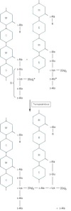
B Lactams usually block the enzyme that ties these pieces together.

With such a wide range of activities against so many organisms one might wonder how they work.  Like all poisons, penicillins inhibit critical life processes in the species attacked. In this case, they inhibit bacterial growth by stopping the repair and replacement of their cell walls.  In affected strains, the cell walls are composed of long chains of six carbon sugars with amino acid side chains that weave the chains together like a net.  The long chains are made by polymerizing N-acetylglucosamine and N-acetlymuramic acid sugars into alternating units.  The side chains are made of repeating amino acid units, which differ per species, but for susceptible species they end with the combination D-alanyl-D-alanine amino acids.  B-Lactam antibiotics are natural analogues of these amino acids, which is to say they can fit into some of the same places as the natural bacterial amino acid piece can.  The antibiotics attach themselves to a special protein (the aptly named ‘penicillin binding protein’) that transports and tethers the D-alanyl-D-alanine amino acids to other chains in the cell wall, blocking its activity(S).  With these proteins blocked the cell wall can’t be replaced or repaired. Lysis results when the cell wall is unable to withstand the osmotic pressure upon it.

It is important to point out that these antibiotics are most active when bacteria are growing and synthesizing cell walls, which is fine for fast growing colonies, like the streptococci.  However mycobacteria, which are famous for causing tuberculosis and leprosy, have a dormant states where they don’t actively synthesize new cell wall components as well as impregnate them with certain lipids that impede the entry of penicillins.  In this context, an attack of this kind would be useless or at the very most a stop gap measure.  Since the colony isn’t completely killed off during treatment, this situation is favorable for the development of antibiotic resistances and the selection of hardy strains of the bacteria.  In a clinical setting these factors complicate issues, with treatments for tuberculosis taking upwards of a year or more to complete.

In a colony of 10^6 bacteria, the probability is that at least one will have resistance to a given antibiotic.  A typical mycobacterial colony will have something around 10^8 members, making the selection of strong, antibiotic resistant strains almost a certainty.  This problem is further compounded by a form of bacterial communication called horizontal gene transfer or plasmid exchange.  This is where a circular strand of DNA, typically encoding a gene producing a resistance, is absorbed by other bacteria allowing the technical means of resistance to spread. This is how bacteria such as the dreaded MRSA have developed and why infections from them are most common in hospitals. In the case of tuberculosis, multi-drug therapies utilizing non-B-lactam antibiotic combinations, like isonaizid-rifampin, can cure 95-98% of cases (S).  The probability that one will have a resistance against both antibiotics is very low and using two will usually kill the bacteria that have a selected immunity to one antibiotic alone.  The other 2-3% typically require specialized treatment and top of the line antibiotics, like moxifloxacin or rifabutin.  However, moxifloxacin resistant strains have been recently identified as a cause for concern (S).

Bacterial resistances usually present via four different routes: Reduced membrane permeability to the drug, production of an enzyme that deactivates the antibiotic (a B-lactamase, for example), a mutation in the PBP site where the antibiotic binds and finally, the creation of a means for drug removal (S).  With penicillins, B-Lactamase production is the most common form of resistance, some of which are highly specific to only one kind of drug (S).  B-lactamases attack the core structure of the penicillin molecule, breaking a critical carbon-nitrogen bond.  Mutations in penicillin binding proteins alter the shape of the binding site, making it harder for the B-lactam structure to fit.  In theory mutations could be great enough to block the penicillin from binding completely.  In practice, however, mutations that cause a PBP to be blocked entirely are often less useful functionally, because these will also block the D-alanyl-D-alanine amino acids they were originally intended for.  Small mutations are more useful to inhibit rather than block the penicillin and keep them from reaching a concentration necessary to inhibit bacterial growth. (S)  Gram negative bacteria usually develop resistances to antibiotic entry by changing the shape of receptors that transport the antibiotic across its membrane.  If the drug can’t pass through the membrane, it can’t attack the cell wall manufacturing machinery.  Another means that changes the membrane permeability is the creation of what is called an efflux pump.  These work to transport the antibiotics back across the membrane after entry.  These are of varied efficiencies, but always work to increase the amount of antibiotic required for efficacy.  Both mutation and changes in permeability are often paired with a B-Lactamase enzyme, making an effective, multi-front strategy for resistance.

Even though we have produced effective weapons against pathogenic bacteria, they have begun to fight back.  They have been largely successful in this regard, calling into question how long the usefulness of these drugs can be maintained.  This has spurred the demand for newer drugs to combat the potential ‘super-bugs’ on the horizon and limited use policies to maintain drug effectiveness in the future.  All in all, penicillin is generally considered non-toxic.  The worst adverse reactions are due to hypersensitivities.  Allergic reactions occur, however, this is a rarity occurring in less than 1% of people and is usually not life threatening (a rash, fever, etc).  Even less have a serious reaction, like anaphylactic shock (0.05% of patients).  In a worst-case scenario, though, desensitization to the drug can be used to effectively treat a given disease.

One thing is for sure is that we will continue to look in awe at the complexity life has to offer.  We hope that new attempts to cultivate and extract new antibiotics will be successful in the future.

{sources available upon request}

To live and function the human body needs energy, the majority of which is produced by the electron transport chain.  The end products from these enzyme catalyzed reactions are oxidized by the oxygen that you breathe and converted into water.  At the center of this complicated process is an incredible  protein that selectively transports oxygen to tissues in need and releases it on demand.  Hemoglobin is a protein carried by red blood cells that has selective affinity for iron, a metal critical to proper function of the body.  The importance of this protein is exemplified by what happens when it is broken.  Sickle Cell Anemia, for example, is caused by a mutation in the gene that codes for the protein.   In this case, the change of a single amino acid (glutamic acid to valine) damages its functionality such that the average life expectancies of people with this disease are only 42 years in males and 48 in women (S).  During this time, those afflicted also experience a host of quality of life maligning health problems, like chronic pain, increased risk of infection and heart disease and vaso-occlusive crises that occur when the misshapen blood cells block blood vessels.

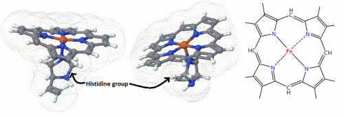
Considering the structure, hemoglobin is a large protein made of four polypeptide chains, sewn together into a tetramer.  There are 2 β chains and two α chains, each of which having a net-like porphyrin ring system.  In human red blood cells, an iron atom is placed in the center of the ring structure, creating a coordinated system called a heme group.  The iron atom is the star of the system and makes the binding of oxygen possible.  It is in the +2 state and has octahedral symmetry, allowing 6 ligands to bind.  The 4 nitrogen atoms of the ring bond equatorially holding the Fe+2 in place, while from the bottom, a histadine amino acid from the protein super structure locks the Fe+2 atom in place.  The open top position is reserved for O2 binding, but in its absence a molecule of water usually takes its place in normal circumstances.

3D images showing the positions of the porphyrin ring, iron and histidine in the heme group. Nitrogen atoms are in blue, iron in red and carbon in the usual grey-black. (3D images courtesy of http://www.3dchem.com By all means check out the structures for yourself using their tremendously useful java based web tool!)

This same system is common throughout other enzymes and respiratory systems.  In humans, Cytochrome C  of the electron transport chain has a similar structure, except that the 6th (top) ligand is a methionine group.  The protein in this case is designed to transport electrons rather than oxygen molecules.  Myoglobin, which is found in muscles and also used for O2 transport, is structurally different, but utilizes a single porphyrin ring system, rather than four.  Enzymes, such as catalase and peroxidase,  also contain Fe manipulating systems similar to the above.  In other kinds of organisms too, the porphyrin ring is utilized in a variety of situations and complexed to many different metals depending on the conditions and needs of the creature involved.  Photosynthetic plants, for example, utilize the same ring, but the metal is magnesium.  Further, some bacteria are also known to use copper as the porphyrin metal of choice.

To make full advantage of this heme system, the binding of O2 in red blood cells is facilitated by a buffer system.  The concentration of any of the components of it increase or decrease the binding affinity of O2 at the hemoglobin binding site.  Proton (H+), CO2, Cl-, and 2,3-Bisphosphoglycerate (BPG) concentrations all have a role in the binding and release of O2 from hemoglobin.  For example, in tissues where the pH (H+ concentration) is acidic and the partial pressure of CO2 is high, the binding affinity of O2 at the binding site will be lowered and will induce hemoglobin to release its contents.  In the lungs, however, the O2 concentration is high compared to that of CO2, facilitating O2 binding.  Normally, BPG is found in equal amounts to hemoglobin, but in situations where O2 is in short supply this balance is undone by the increased production of BPG.  With BPG, binding to one of the active sites reduces the O2 binding affinity by about 25 times, thus when BPG concentration is high it pushes for a release of O2 into tissues that need it.

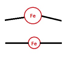
The larger Fe staggers the planar porphyrin ring, where as the smaller version fits better, contributing to stability.

Aside from outside effects promoting binding, there are effects coming essentially from the design of the system itself that have to be considered as well.  The question of why iron and not some other transition metal is of special relevance here.  Using iron as the central metal, in this case, yields benefits in terms of performance and binding specificity to oxygen.  Cobalt, for instance, is used in similar systems like those of vitamin B12’s corrin ring, but while both cobalt and iron contain d orbitals that could bind, only iron allows for the perfect balance between size and binding specificity in this system.  While unbound, the Fe+2 atom is just a little too large to fit into the normally planar porphyrin ring.  In this case, it is in a high spin state where the molecular orbitals are further away from the atom, giving it a larger size.  The high spin state is larger, because the Pauli exclusion principle prohibits the atom’s 3d electrons from getting too close to one another due to repulsions.  Thus, additional space is needed to house the electrons in orbitals further away.  However, when the Fe+2 binds to an oxygen molecule the electrons can be shuffled into orbitals that are closer to the atom.  Its orbital symmetry changes to a low spin symmetry and CLICK ! the Fe+2 shrinks just enough to fit snugly into the porphyrin ring system.

A silly, but useful comparison.

This part also contributes to a critically important finding: that the binding of O2 is cooperative, in that the binding of one O2 molecule will facilitate the binding of another until all four spaces are filled.  It has been experimentally determined that the binding energies (the Ka) are increasingly smaller for each molecule of O2 bound to hemoglobin.  This happens, in part, because of the protein’s structure.  When the O2 binds and the Fe+2 clicks into place, it pulls on the histadine residue below it, stretching the other protein superstructure bonds.  This pulls slightly on the other 3 Fe+2-histadine bonds.  Much like those dancing string toys, pulling on the string at the bottom causes the toy’s arms and legs to move in a concerted action, which is similar in a way to the physical reaction of the other binding sites.  The additional pulling on the other histadine residues facilitates binding of the other three, such that the binding energy is progressively reduced with each binding until all four slots are filled.

Other structural contributions also play a large role in binding specificity.  Above the plane of the porphyrin molecule lies another histadine residue that physically blocks the strongest and most effective bonding interactions from occurring.  Since the Fe+2 atom is locked into place, the best bonding interactions would come from ones that provide the most overlap of their molecular orbitals, which are those that are end-to-end.  However, with the histadine in the way, these are prevented from occurring.  This is a good thing, because strong covalent interactions in enzymatic reactions, like those seen in the binding of carbon monoxide (CO) for instance, are usually toxic and are difficult to break under normal conditions.  For head-to-head binding to CO, the interaction is estimated to approximately 1000 times as strong as those between O2, illustrating its toxic potential.  In this case, the CO-Fe binding is still strong, but not so much as to completely block removal.  People who have suffered CO inhalation are often given pure oxygen in an attempt to out pace CO binding and ensure that the person continues to have a supply of oxygen.  This hindered binding is also helpful in normal activity as well, since the binding symmetry to O2 is bent as well it further facilitates it’s release into tissues in need.

Yes, it’s about iron again, but it’s quite interesting stuff.

{sources available upon request}

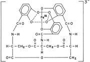
Enterobactin

A little over 40 years ago it was discovered that bacteria utilize a class of metal chelators called siderophores to capture iron from their environments.  Iron is an important element for the growth and development of most kinds of bacteria.  It is used as a critical element in electron transport chains for energy production, the removal of dangerous reactive oxygen species and the functional element in the reactive sites of many important enzymes.  The finding was critically important to the field of medicine, as the iron scavenging activities of pathogenic bacteria are most often detrimental to the host.  Even though humans and other animals have developed sophisticated systems to sequester iron and make the body inhospitable to these bacteria, likewise they have developed a variety of means of out-competing their hosts for the valuable element.  Neisseria gonorrheae and N. meningitidis, for example, have evolved the ability to directly capture iron containing lactoferrin proteins from their human hosts, a testament to our continued co-evolution (1).  Still others have relied upon producing their own iron scavenging siderophores to fish out this valuable metal from fluid and protein sources.  Escherichia coli , for example, are known for producing enterobactin and have one of the most effective iron scavenging systems known, which at biological pH easily out competes endogenous defenses for iron.

Recently, it has been noted that these compounds are of critical importance to bacterial growth, especially in the ultra-low iron concentrations of bodily tissues.  Rather than tighten their belts in the toughest of times, some kinds of pathogenic bacteria turn on

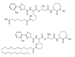
'Like dissolves like': different kinds of Mycobactin are used in different mediums. The long aliphatic chain endows fat solubility to the molecule, allowing it to shuttle through the waxy mycobacterial membrane easily.

these scavenging systems to forcibly acquire the iron needed for growth.  Mycobacterium tuberculosis produces several siderophores, called mycobactins, that are medium specific in their activities.  The two differ by a water soluble or fat soluble group attached to the siderophore skeleton.  Water-soluble mycobactin T is released into aqueous mediums, whereas fat soluble mycobactin T is designed to take pirated iron across the waxy mycobacterial cell membrane (2).  When iron is in good supply, however, production of these is unnecessary and is down-regulated to conserve valuable energy and resources.  Below the level of production of these siderophores, however, lies a regulated system of control.

Generally siderophore production is controlled by gram negative bacteria’s “Fur” and gram positive’s “DtxR” systems, which have  transcription elements that are bound by iron.  Thus, when intracellular iron concentrations fall below a threshold (approximately 10^-6 M for many pathogenic bacteria) the iron lynchpin holding back the transcription element is gone, activating the siderophore production system.  In 1999, researchers at the University of Queensland showed that Mycobacteria require the production of siderophores for growth in human tissues and macrophages (2).  They used genetic engineering techniques to remove the instructions for the enzyme MbtB, which is a critical component of mycobactin synthesis.  MtbB effects the final step in mycobactin synthesis, the coupling of a salicylic acid group to the siderophore’s finished structure.  After creating an MtbB knockout strain, its ability to produce siderophores was tested by an assay that measured the absorbance of the growth medium.  They used a colored dye compound that has a known affinity for iron in solution.  If a bacteria were to produce something that has a higher affinity for iron than the dye, this would be measured as a drop in absorbance, as the siderophore would compete with the dye for the iron.  They found that after comparing these bacteria against wild type strains that they were unable to produce siderophores and the deficient strains experienced retarded growth both in plate medium and in live macrophages (in vivo vs. in vitro tests).

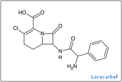
The understanding of how important these chemicals are for bacterial growth has led to renewed attempts to develop antibiotics against them.  In the late 80 and early 90’s the ‘trojan horse’ tactic of using siderophore-antibiotic conjugates and piggybacking them into a cell began to bear fruit with successes being discovered in a wide variety human diseases (3)(4)(5).  One group of scientists developed conjugates using the carbacephem antibiotic, loracarbef.  They found that the mixed catecholate-hydroximate siderophore they used gave the conjugate compound 2,000 times more potency than the parent drug alone (6).  Though resistances to this tactic still developed, these are thought to leave the bacteria at a disadvantage, because the mutations hinder the siderophore uptake system rather than attack the antibiotics themselves.  This limits access to a vital nutrient and leaves the mutants more susceptible to iron starvation and hindered growth.

Though the trojan horse tactic has yielded positive results, there are opportunities for even greater exploitation.  Techniques involving small molecules that attack that actual production of siderophores could provide another avenue to beneficial therapy for these diseases.  These drugs, much like statins that block the enzyme HMG-coA, would block a critical piece of the siderophore production pathway.  In the particular case of mycobactins, the final step in biosynthesis is the attachment of a salicylate group to the mycobactin skeleton.  Researchers at Cornell published results in 2005 showing that inhibitors of the enzymes that accomplish this task are potent inhibitors of M. tuberculosis and Yersinia pestis (7).  They synthesized a compound, SAL-AMS, that closely resembled a reaction intermediate and measured its effect upon the growth of bacterial cultures in mediums of low iron concentration.  They found that it successfully inhibited the enzyme and drastically reduced bacterial growth in the cultures.  It was shown to have an IC50(*) of 2.2 ± 0.3 μM for M. tuberculosis and about 51.2 ± 4.7 μM for Y. pestis in an iron limited medium.  Though in mediums with high concentration of iron the chemical was ineffective against Y. pestis, but it was found that the chemical might have unknown inhibitory properties against M. tuberculosis, as:

“Salicyl-AMS (tested at up to 8 X IC 50) was not active against Y. pestis in iron-supplemented medium, in which siderophore production is not required for growth.  Under these conditions, salicyl-AMS (tested at up to 180 x IC50) did inhibit M. tuberculosis growth, albeit with an 18-fold increase in IC50 (39.9 ± 7.6 μM).  This suggests that, in addition to blocking siderophore biosynthesis, salicyl-AMS may also inhibit M. tuberculosis growth by other mechanisms.”

Furthermore, researchers at the university of Minnesota developed similar nucleoside inhibitors of MbtA, one of which ” rivals the first-line antitubercular isoniazid” in activity against the bacteria (8).  This tactic follows the same reasoning as SAL-AMP above, as the nucleoside inhibitors attack the same pathway to inhibit siderophore end production.  The research was centered around making logical functional modifications to known structures of inhibitors and choosing the most effective ones for further testing.

Comparison of the pathway intermediate, the Cornell inhibitor and one of the Minnesota nucleoside inhibitors.

These findings are coming just in the nick of time it seems, as certain drug resistant strains of M. tuberculosis have become big news recently.  Extensively drug resistant tuberculosis is a kind of tuberculosis that is resistant to at least two of the top line drugs used to normally treat it (typically isoniazid and/or rifampicin) and a member of the quinolone antibiotics (ciprofloxacin).  Tuberculosis is generally a challenge to treat in the first place, with treatments typically taking up to a year or more to complete.  The loss of the first line drugs and reliance upon second line increases the risk of side effects and patient noncompliance to the already long course of therapy.    This can further complicate the issue, as it could lead to the obsolescence of the few active  drugs used to treat the disease, because resistances to one drug are usually useful against the whole family of drugs.

β-lactamases typically attack the carbonyl in the β-lactam structure, destroying the ring. Nafcillin has a large group that hinders these enzymes from getting too close.

An example of this can be seen in bacteria that produce β-lactamases, as these strains are often cross-resistant to all unprotected β-lactam antibiotics.  Some  penicillins have been designed with bulky groups attached to the skeleton in an attempt to hinder these enzymes.  Nafcillin, with its large 2-ethoxy-1-naphthoyl group, is very effective at blocking these enzymes for the most part.  However, even this tactic has its limits as methicillin resistant Staphylococcus aureus (MRSA) and oxacillin resistant Staphylococcus aureus (ORSA), both have developed resistances against these drugs such that, “From 1999 through 2005, the estimated number of S. aureus–related hospitalizations increased 62%, from 294,570 to 477,927 (9),” and “MRSA accounts for an estimated 12% of all nosocomial bacteremias, 28% of surgical wound infections, and 21% of nosocomial skin infections.  Infections secondary to MRSA result in excess costs of approximately $4,000 per patient per hospitalization compared with patients infected with methicillin-susceptible S aureus (MSSA)”(10)(11).

Since the discovery of penicillin, Alexander Fleming hypothesized that bacteria would develop resistances to the antibiotics used to treat them.  Now, more than ever, the development of new antibiotics must be pursued.  Human defenses have always provoked a counter-response from our pathogens, the most successful tactic selected out.  Much like the pathogens that infect us, we must ‘evolve’ a newer understanding of bacterial biology, which will offer us a foothold to better, more effective treatments.

——

(1) Genetics and Molecular Biology of Siderophore-Mediated Iron Transport in Bacteria

JORGE H. CROSA

(2) The salicylate-derived mycobactin siderophores of Mycobacterium tuberculosis are essential for growth in macrophages

James J. De Voss, Kerry Rutter, Benjamin G. Schroeder, Hua Su, YaQi Zhu, and Clifton E. Barry III

(3) Design, Synthesis, and Study of a Mycobactin−Artemisinin Conjugate That Has Selective and Potent Activity against Tuberculosis and Malaria

Marvin J. Miller, Andrew J. Walz, Helen Zhu, Chunrui Wu,Garrett Moraski, Ute MÖllmann, Esther M. Tristani, Alvin L. Crumbliss, Michael T. Ferdig, Lisa Checkley, Rachel L. Edwards, and Helena I. Boshoff

(4) Species Selectivity of New Siderophore-Drug Conjugates That Use Specific Iron Uptake for Entry into Bacteria

M. S. DIARRA, M. C. LAVOIE, M. JACQUES, I. DARWISH, E. K. DOLENCE, J. A. DOLENCE, A. GHOSH, M. GHOSH, M. J. MILLER,    AND F. MALOUIN

(5) Siderophore-Based Iron Acquisition and Pathogen Control

Miethke M, Marahiel MA.

(6) Iron Transport-Mediated Antibacterial Activity of and Development of Resistance to Hydroxamate and Catechol Siderophore-   Carbacephalosporin Conjugates

ALBERT A. MINNICK, JULIA A. McKEE, E. KURT DOLENCE, AND MARVIN J. MILLER

(7) Small-molecule inhibition of siderophore biosynthesis in Mycobacterium tuberculosis and Yersinia pestis

Julian A Ferreras, Jae-Sang Ryu, Federico Di Lello, Derek S Tan & Luis E N Quadri

(8) Antitubercular Nucleosides That Inhibit Siderophore Biosynthesis: SAR of the Glycosyl Domain

Ravindranadh V. Somu, Daniel J. Wilson, Eric M. Bennett, Helena I. Boshoff, Laura Celia, Brian J. Beck, Clifton E. Barry, III, and Courtney C. Aldrich

(9) Hospitalizations and Deaths Caused by Methicillin-Resistant Staphylococcus aureus, United States, 1999–2005

Eili Klein, David L. Smith, and Ramanan Laxminarayan

(10) Baquero F. Gram-positive resistance: Challenge for the development of new antibiotics. J Antimicrob Chemother. 1997;39(suppl A):1–6.

(11) Kopp BJ, Nix DE, Armstrong EP. Clinical and economic analysis of methicillin-susceptible and -resistant Staphylococcus aureus infections. Ann Pharmacother. 2004;38:1377–1382.

(*) The IC 50 is the half maximum inhibitory concentration of antibiotics, a typical measure of effectiveness of the drug.

Gyoza are a Japanese rendition of an old Chinese recipe for pork dumplings. This is one of the more ubiquitous things Japanese families serve and are generally found in most restaurants, especially ramen stops and donburi places.  Some restaurants, like “Gyoza no Oo” (‘king of gyoza’), specialize in them.  These are so ubiquitous that grocery stores usually stock a manufactured freeze dried version.  Generally, they are served as a side dish or even a main course where a family will eat off of a large hot plate in a similar fashion to yakiniku.

The recipe is fairly easy and most of the ingredients can be found in a standard Asian foods grocery store.  The general ingredients are round gyoza wrappers, ground pork, garlic, nira (similar to chives or green onion), shredded cabbage, soy sauce, sake, ground fresh ginger and some black pepper for basic flavoring.  Additionally, we like to add some red miso paste, a little yellow onion and ground beef

This is the meat mix.

so that the meat is a 30/70 mix.  The miso paste is also something that we just like; it gives them an earthier, sweet taste.  Some recipes also add sesame oil to the mix, but I’m not really fond of it personally.  There are many locally accepted additions to this framework that are typically regionally famous.  For example, I’ve had crunchy renkon (lotus root) gyoza before, which was interesting as a local oddity.

Lets get down to it. Boil all vegetables until soft then cut and drain most of the excess water. Mix the pork, spices and a heaping spoon of miso paste.  Add a spoon of soy sauce and a spoon of sake.  These two ingredients and the pepper are generally to personal taste.  Cover the meat and let it stand for 10 minutes or so.

Next comes the tedious wrapping process:

The first thing you need to know is how to work with dumpling wrappers.  Dumpling wrappers work best when they are cold and usually people buy them in the store’s freezer section.  Let these thaw either in a fridge or outside to near room temperature. The key is to not let them get too warm or they’ll melt and stick together.  Once that happens there’s nothing you can really do with them.  Usually, they’re coated in starch to aid in the wrapping process by removing that extra moisture that’s always around in these climates.  The biggest point is to not let them get them wet before using them.  Take a small ball of meat and put it on the center of the wrapper then while in the palm of your hand line the edge of the wrapper with a little bit of water.  This will make it stick when you fold the wrapper up. Folding it is hard to describe, but pinch the outside edge and fold it over about 1 cm and repeat this 4-5 times. Set aside somewhere dry and wait to cook. Repeat this process several hundred times and you’re set for dinner.

To cook them place them in a fry pan and sear the bottom until golden brown.  This will help them release from the pan and give them a crispy skin once their done.  Once they’re browned, add just enough water to cover the bottom of the pan.  Cover with a lid and listen to the sound.  When they’re done, you’ll hear the sound of the water boiling change to a higher note.  This means that most of the water should be boiled off and the gyoza should be ready to eat.  Cut the heat and enjoy.  We eat them right off the hot plate.  Another option aside from frying them would be to use them in soups, in a similar way to wonton soup in Chinese restaurants in the states.

One more thing, I almost forgot the sauce. People usually dip them in some kind of sauce, variations of which are almost endless.  You can buy them or make them from stuff in you kitchen.  Most of these sauces have a soy sauce base, with something else to give it a little spice.  I’ve used ponzu (a kind of citrus soy sauce), raiyu (red chill oil) or just delicious sriracha.  Some people also add sesame oil to the mix, but I’m not a fan.

Lonely prey, stalked, waits to be eaten

Polyethers are made by the anionic polymerization of an epoxide and a strong base like an alkoxide ion. This reaction polymerizes the two compounds and creates repeating ether (R-O-R) links between them making long chain compounds from 300 g/mol to 10,000,000 g/mol. This kind of reaction is ended when an alcohol like methanol deactivates the end chain anion by donating a hydrogen atom. As for physical properties, they have a high solubility in water because of their ability to make hydrogen bonds. Industrially they are marketed under many names, but a carbowax (varied types) seems to be the most popular name.

These chemicals have a surprising amount of uses as surfactants in industry, including foods (filler: probably McD’s), cosmetics and pharmaceutics; in biomedicine, as dispersing agents (timed release medicines), solvents, ointment (the lube, dude: KY) and suppository bases (GoLYTELY, GlycoLax), vehicles (car waxes), new types of body armor (liquid sheer thinking fluid body armors), cloth and fabrics (think spandex), tablet excipients (inactive ingredients in vitamins medicines). They have a low toxicity (1) and are finding new use in the repair of damaged nerve cells in animal trials (2), as well as use as a colo-rectal cancer preventative.

Unfortunately, the human trials are too late for Christopher Reeve and others who suffer a similar fate, but perhaps they will happen sooner than we think and hopefully (if we’re lucky the research is applicable to humans) many will reap the rewards of this research. Trials have been conducted with guinea pigs, dogs and such with fairly encouraging results (2), but the effect on human damage is unknown. One thing is certain: its definitely not an ordinary laxative.

1. Toxicology

http://www.mindfully.org/Plastic/Pol…ycols-PEGs.htm

2. Animal tests:

http://www3.interscience.wiley.com/c…TRY=1&SRETRY=0
http://www3.interscience.wiley.com/c…06760/ABSTRACT
though not applicable to humans, this link talks about its use as a colorectal cancer preventative:
http://www.inra.fr/reseau-nacre/sci-…t/indexan.html

3. Nerve guides help regeneration

http://www3.interscience.wiley.com/c…97042/ABSTRACT
http://www3.interscience.wiley.com/c…32586/ABSTRACT

4. general interest news

http://seattlepi.nwsource.com/health…_spinal04.html

As I was running yesterday a thought occurred to me as the steady, percussive hum of wings buzzed by me several times: one giant Asian hornet (an osuzumebachi) is startling, but several dozen is pretty scary. Apparently, I had run too close to a nest and had to hot foot it out; I was lucky.

These things are pretty amazing creatures, with an average body length of 2 inches and a wing span of 3, you will stand up and take notice when they fly past. As with many bee and hornet stings single stings can be deadly as a result of anaphylactic shock, but this is fortunately a rare occurrence. Nonetheless, the hornets are nothing to take lightly as the venom contains an interesting mix of nastiness, as well as a barbless stinger, which it can use repeatedly unlike regular honey bees. The venom contains neurotransmitters like serotonin and acetylcholine, which is responsible for pain transmission, a neurotoxin called mandaratoxin, tissue dissolving enzymes such as phospholipase-B, a mast cell degranulating peptide called mastoparan and a pheromone that instigates other hornets to swarm and attack. Aside from pain, the stings usually leave a dime-sized scar not unlike those of certain poisonous spiders in the southwestern US.

The hornets terrorize smaller, weaker prey like praying mantii and Japanese honey bees. The bee’s stingers are too small to penetrate the hornet’s tough, armored exoskeleton and thus have little defense from a full on attack. They reputably work with ruthless efficiency, a single hornet able to kill as many as 40 bees per minute and a few hornets only a few hours to slaughter a 30,000 bee colony. Interestingly, the bees have a small defense against them in their greater tolerance for heat. Typically, the bees will entice a scout inside the hive, where they will attempt to smother him in a bull rush. Once he is covered, the bees will vibrate their flight muscles increasing the heat in the area to around 47C. The hornets can’t survive past 45C, preventing the scout from calling reinforcements.

Link: http://www.youtube.com/watch?v=JDSf3Kshq1M
Link: http://www.youtube.com/watch?v=hpcHH1EpTZM

Even though, these may seem like kings of the forest, human initiative has once again found ways to make use of nature. The Japanese love energy drinks, one of the more famous products, VAAM is of particular note. As advertising shows it has been the favorite of many a marathon runner and endurance athlete. Yet a little less advertised fact is that it’s a chemical copy of the stomach contents of these hornets. More specifically, it is a copy of the secretions of the colony’s larvae, which the adults feed upon as they lack the ability to digest raw protein themselves. How’s that for wild? This mixture of predigested amino acids and sugars supposedly allows them to fly distances of 60 miles per day at rates of 25-40 miles per hour.

Sounds perfect for runners, now lets have a toast!

As gross as that sounds many professional athletes here swear by its supposed benefits and from personal experience it tastes pretty good. The question of the day, though, is one of efficacy: Does it work? Dr. Takashi Abe of the Institute of Physical and Chemical Research in Japan seems to think so. “VAAM works by helping the body burn the energy it stores more efficiently”…”[It] expedites the metabolism of fat and promotes better hydration,” said Dr. Abe. Animal studies conducted by Abe concluded that mice that were fed VAAM could swim twice as long as those that were fed only water and 25% longer than those fed casein, a protein found in milk. Their blood also contained fewer fatty acids than those of the other groups, an indication of how efficiently the mice were burning their fat reserves. Details are sketchy as to how it might actually accomplish these feats. One possible route may be through the branched chain amino acids in it, which increase the effect of insulin in the body. The product contains a mixture of leucine, isoleucine and valine, of which leucine is considered the most effective at this. Leucine reportedly “increases insulin output by 221%,” if taken with carbohydrates after exercise.

If you’re thinking: “At last the perfect supplement to my admittedly weak dietary habits, that will propel me to epic heights of fame and fortune,” then you’re most likely going to be disappointed (mostly in yourself). I’ll save you the trouble and clue you in to something important: the supplement industry is mostly a joke played on the lazy for the fun of a few bored chemists. Though there might be a slight benefit to some supplementation, there is no substitute for hard work, but we delight in trying to make you think there is. (Next year’s big craze will be whale sperm marketed under the name SPEaRM—->.) On the other hand, if you’re looking to try something that tastes good to replenish your vital fluids during or after a workout, then this might be up your alley.