Feeds:
Posts
Comments

Archive for the ‘Chemistry’ Category

Muscular dystrophies are a class of degenerative diseases characterized by progressive muscular weakening, myopathy and paralysis.  Among these, one of the most famous is Duchenne muscular dystrophy (DMD), which affects about 1 in every 3000 males born in the United States.  This class of diseases faces the very limits of medical therapy, as there are currently no clinical treatments available to halt the progression of symptoms.  With only supportive care available, patients are left to progressively worsen, typically ending with loss of physical mobility, the ability to breathe independently and usually fatal heart related complications.  The disease maligns quality and length of life, such that most only live into their twenties and are confined to a wheelchair around their teens.  Only in very rare circumstances have any lived into their 30’s or 40’s.

It wasn’t until fairly recently that an accurate understanding of the pathology involved became clearer.  With DMD, mutations from reading frame shifts in the gene producing the dystrophin protein were found to be the root of the problem.  Dystrophin is a critical protein involved in muscle structural support and intracellular signaling with the extra-cellular matrix.  For any kind of contraction, a muscle must have an anchor and this protein helps play this part by linking the strand to a support structure called the DAP complex, which in turn links to the extracellular matrix.  Like a car without shock absorbers, muscle cells are more susceptible to physical membrane damage and slowly whither under the stress.  Damaged, leaky membranes are a classic indicator of failed cell viability and seen as the hallmark of dying cells, a fact corroborated by typical diagnostic staining procedures using cell impermeable dyes.  Dystrophin is also heavily involved with intracellular signaling from responses outside the cell.  Generally, proper signaling is necessary for continued cell viability.  Without it cells are usually marked for termination by immune system cells like macrophages, which seek out this kind of aberrant signaling.  DMD affected muscles are often infiltrated by these kinds of cells, which further increase the damage by initiation of an inflammatory response.

One of the hallmarks of this disease can be seen in slides of affected muscle.  Healthy cells are ensheathed by a thin membrane called the endomysium, but in diseased tissue the spaces around the muscle cells are filled increasingly with fat or fiber-like cartilaginous tissue.  This is called endomysial fibrosis.  As macrophages realize something is wrong, they infiltrate the area and initiate an immune response causing inflammation.  This further weakens the damaged cells and whittles the number of them down slowly, which accounts for the progressive weakening pathology of the disease.  The dead cells are typically replaced by this white tissue matrix.

Many hypothesized early on that gene therapy held the greatest promise to cure this disease.  After all, in order to truly cure a disease of genes, you must fix those broken genes.  Though pharmaceutical treatments reducing immune system response to the disease may improve conditions temporarily, they most likely won’t offer the long-term increase in survivability sought after for so long.  As such, many new methods and techniques have been proposed as models for new gene therapy based strategies.  Many of these have proven successful in mouse models and a few have even had some success in small clinical trials.  These cover a wide range of areas and strategies, such as using viral vectors for gene transplantation, up-regulation of suitable endogenous genes and forced expression of tailored mini genes.  Each of these has found success as a possible route towards a therapy, but each with its own drawbacks and limitations.  The master stroke, it seems, is still some time off, but even closer and within our child-like grasp at the cookie jar.

  1. Viral vectors and transgene infection

A now famous example comes from researchers at the University of London.  They worked with viral vectors to introduce a dystrophin replacement gene into muscular dystrophic mice.  Viral vectors are complicated, because they can provoke a response from the immune system, can possibly cause disease and have a limited storage capacity for any potential gene transplant.  They found a balance with the virus called Adeno-associated virus (AAV).  These viruses are able to infect many kinds of cells, can be produced without their endogenous viral genes, have never been shown to cause a human disease and also require a helper virus to replicate themselves after infection.  This combination of characteristics limits the dangers associated with infection to a degree and makes them good candidates for use in human gene therapies.

One of the main problems they faced was the small storage capacity the virus could carry.  The dystrophin gene is one of the largest known (2.4 megabases), so they had to make some kind of compromise.  They worked around this by manufacturing a mini-gene that was much smaller than the actual gene that coded for the dystrophin protein.  This was made possible by knowledge of the genomes of those affected by the disease.  A close cousin of Duchenne muscular dystrophy (DMD) is called Becker muscular dystrophy (BMD) and is characterized by a lesser severity of symptoms.  Surprisingly, the gene mutations in BMD, though sometimes ‘affecting ~ 50% of the gene itself’, affect much less critical locations than mutations in DMD.  Mapping these locations allowed them to whittle out less critical pieces of the DMD gene.  They developed a mini-gene that would produce a lesser functional, but still useful dystrophin protein, which was still able to fit within the size constraints of the viral carrier.

Once the mini-gene was completed they paired it with a transcription promoter from the cytomegalovirus to force gene transcription and production of the protein once the virus infected a cell.  They injected the virus into mice expressing a DMD phenotype and found that the mini-gene was successfully expressed in “greater than 50% of the muscle fibers 20 weeks after infection”(S).   It was found that it relieved aspects of DMD pathology, particularly rebuilding the DAP complex and improved muscle structure.  This culminated with mice test groups showing characteristics of a lesser severe form of the disease.  Furthermore, experiments showed that even with a low 20-30% level of gene expression, there was a substantial reduction in DMD pathology overall with the use of this mini gene.

2.       Increasing transcription of gene with artificial constructs

As it turns out the dystrophin protein is not the only one utilized to link the muscle to the extracellular network.  Before birth, another similar protein called utrophin is used preferentially for the same purpose, but is replaced almost entirely by dystrophin after birth. The protein exhibits 80% similarity with dystrophin and remains expressed during the course of the disease.  Researchers at the Istituto di Neurobiologia e Medicina Moleculare in Rome surmised that if preferential expression of utrophin was re-established it might provide a route to a cure or a reduction of symptoms to make it manageable.  Using an artificial transcriptional element called “Jazz”, they were able to restore muscle integrity and prevent the development of DMD in mice test groups.

It has long been known that the sudden expression of new, ‘foreign’ proteins runs the risk of causing an immune response as immune cells target these new proteins as antigens.  So just giving a person an extra supply of dystrophin won’t work as a treatment for the disease.  It was found that when a cell lacks proper functioning dystrophin, it up regulates utrophin to compensate, but the level is insufficient to prevent disease progression.  Since this protein is already expressed increased production could reduce complications caused by immune system interference.  Many previous studies have confirmed that: “Increased expression of utrophin restores plasma membrane integrity and rescues dystrophin-deficient muscle in mdx mice.”(S)

A ‘zinc finger’ is a recognition element that can interact with specific sections of DNA.  In this study, an artificial zinc finger was manufactured to correspond to a section of the promoter in the utrophin gene in both the mice and human genes.  Upon testing, they found that it was able to successfully bind to its specific DNA target sequence and increase production of utrophin expression to 1.8 times that of controls.   This increase in production also translated to a therapeutic benefit in mice test groups, showing increases in muscle size, fiber regeneration and lower serum levels of creatine kinase, a chemical identifier of muscle necrosis.

Possible benefit was further characterized by in vitro testing of muscles using electro stimulation.  Weak muscles, deficient of contractile force are a hallmark of DMD, yet those treated with the ZF ATF “Jazz” showed the opposite in excised diaphragm and extensor digitorum longus muscles.  These were able to perform longer and more sustained contractions, than diseased control groups.  Membrane integrity was also tested by staining with procion orange dye.  This fluorescent dye is usually only taken up into a cell with a leaky membrane, so it is often used to assess membrane integrity.  Sustained contractions to a muscle with a DMD phenotype would cause membrane damage and usually exhibit a greater dyed area than that of a healthy cell under the same conditions.  Muscles tested and stained under these conditions showed positive results for Jazz treated test sets, with greater dyed areas observed in the DMD cells.

3.       Antisense Oligionucleotides:

Antisense oligionucleotides are a class of nucleic acids that have also been tapped as a possible route to a DMD therapy.  In 2008, researchers at Oxford published a study testing the hypothesis that these could induce at least partial dystrophin protein expression by pushing the reading frame over in mutated muscle cells.  The idea in this technique is to use a technique called ‘exon skipping’ to push the ‘out of reading frame’ portions of the protein back into the reading frame and produce a partially functional, “becker-like” dystrophin protein.  This technique was shown to have benefit in not only mice, but also in humans in a proof of concept test in 2007.

Up until now, universal changes in expression from AOs were difficult to attain, with high percentage expression limited to skeletal muscle groups, but only limited expression in critical heart and diaphragm muscles.  This was a huge limitation to its possible use as a therapy, as “cardiomyopathy is a significant cause of morbidity and death in DMD patients” (S).  Without a corresponding effect upon heart muscle, any increase in overall muscle dystrophin expression would only exacerbate any heart conditions a DMD patient (or mouse) might have.  However, this new test was different and achieved much more favorable results in mice.  The question is: what did they do different?

It was surmised that the previous tests of AOs had limited entry into exclusive, protected environments like the heart.  They needed a tool that would allow access to these areas without destroying the gains made in earlier tests and this was achieved by conjugating the AOs to an arginine rich peptide scaffold.  Arginine is a positively charged amino acid and its use in the peptide yields an overall positive charge.  These kind of chemicals are thought to use special cell-mediated uptake systems in common with glycosaminoglycans, thus like a Trojan horse they facilitate the entry of whatever cargo they might bring.

Three weeks after injection, “between 25 and 100% of normal dystrophin levels had been restored in body-wide skeletal muscles” and “even in the diaphragm almost 25% of normal dystrophin protein was restored.”  Restoration was also seen in the heart, but not quite as high as that of skeletal muscle: “levels between 10 and 20% of that found in normal mouse heart were typically seen in all western analysis in all treated animals.” These results also corresponded with a function increase in muscle contractility and lower serum creatine kinase levels, both indicators of improved muscle ability and reduced muscle damage.

To build a tower on the sea.

Though these seem like giant steps towards a cure for one of the great diseases of man, there will undoubtedly be more questions than answers.  These are exciting times.

Every one of these people worked really hard. Please read these sources first hand (at least these):

1. “Adeno-associated virus vector gene transfer and sarcolemmal expression of a 144 kDa micro-dystrophin effectively restores the dystrophin-associated protein complex and inhibts myofibre degeneration in nude/mdx mice. Stewart A. Fabb, Dominic J. Wells, Patricia Serpente, george Dickson.  Human Molecular Genetics, 2002, vol. 11, No. 7, pgs 733-741.

2. “Expression of human full-length and minidystrophin in transgenic mdx mice: implications for gene therapy of Duchenne muscular dystrophy.” Wells DJ, Wells KE, Asante EA, Turner G, Sunada Y, Campbell KP, Walsh FS, Dickson G.  Hum Mol Genet. 1995 Aug;4(8):1245-50.

3. “The artificial gene Jazz, a transcriptional regulator of utrophin, corrects the dystrophic pathology in mdx mice.”  Maria Grazia Di Certo, Nicoletta Corbi, Georgios Strimpakos, Annalisa Onori, Siro Luvisetto, Cinzia Severini, Angelo Guglielmotti, Enrico Maria Batassa, Cinzia Pisani, Aristide Floridi, Barbara Benassi, Maurizio Fanciulli, Armando Magrelli, Elisabetta Mattei, and Claudio Passananti. Hum. Mol. Genet. (2010) 19 (5): 752-760.

4. “Cell-penetrating peptide-conjugated antisense oligionucleotides restore systemic muscle and cardiac dystrophin expression and function.”  HaiFang Yin, Hong M. Moulton, Yiqi Seow, Corinne Boyd, Jordan Boutilier, Patrick Iverson and Matthew J.A. Wood.  Hum. Mol. Genet. (2008) 17 (24): 3909-3918.

5. “Matrix metalloproteinase-9 inhibition ameliorates pathogenesis and improves skeletal muscle regeneration in muscular dystrophy.”  Hong Li, Ashwani Mittal. Denys Y. Makonchuk, Shephali Bhatnagar and Ashok Kumar.  Hum. Mol. Genet. (2009) 18 (14): 2584-2598.

Read Full Post »

“Wolf’s liver taken in thin wine, the lard of a sow that has been fed upon grass, or the flesh of a she-ass taken in broth.” –Pliny the Elder

Ring of Inhibition

We’ve come a long way in some ways, at least since “flesh of a she-ass” was considered a specific remedy from disease.  Though, did you know that in early Greece moldy bread was a remedy against infection of wounds?  The mold was presumably a kind of penicillium that produced chemicals inhibiting bacterial growth.  On that fine day in 1928, Alexander Fleming had no idea he would discover something that would change medicine forever.  After coming back from vacation and sifting through the piles of clutter in his office he noticed a culture of staphylococcus in which a blue mold was growing.  The mold had pushed back the staph in a characteristic ring of inhibition.  As the story goes, he hypothesized that the mold must be producing something that retarded bacterial growth.  Penicillium chrysogenum produces the chemical backbone used to produce penicillin, an antibiotic which caused a great leap forward in western medicine.  The effect has been so great that the old killers of yesteryear, the terrible diseases that for ages struck fear in the hearts of man are no longer thought of in the same grave tone and are considered more of a nuisance to public health, a mere trifle in consideration of the far greater concerns of the general populace.  Perhaps the greatest triumph is the reaction of surprise that “people still get that?!”

The back bone of penicillin antibiotics, 6-aminopenicillanic acid, was originally isolated from cultures of Fleming’s Penicillium

Various functional groups can be added to the amino side chain to produce different drugs.

notatum.  It is a secondary metabolic product, released only when the mold is under environmental stress.  Early manufacturing methods were hampered, because finding just the right conditions for the mold to release the chemical proved difficult, thus production was initially low.  When the first human trial in the U.S. began in 1942, doctors used half of the nation’s total production of penicillin to cure him. The medical value placed upon it began a race to massively increase its production. Researchers from both Britan and the US worked tirelessly to not only elucidate the shape of the molecule, but develop better processes for producing it.  In 1943 a nationwide search was held to find better strains from which the B-lactam skeleton could be obtained.  The lucky winner was provided by a moldy cantaloupe from Illinois, which produced many times the amount of penicillin than Flemming’s original.  This strain exceeded expectations and was quickly put to work manufacturing the raw penicillin components.  At the time, it was so prized that during World War II doctors would collect their patient’s urine to isolate and reuse the excreted portion of penicillin.

Today genetically engineered strains manufacture it in large tanks where conditions such as the pH, temperature and diet are all automatically controlled to elicit maximum output (S).  The growth medium is key to the entire process, where the goal is to put them under nutritional strain.  Growth mediums are usually proprietary information, but they typically contain a source of carbon and nitrogen, which are chosen to make them work harder to gain the nutrition they need to survive.  Penicillium notatum prefers the simple 6 carbon sugar glucose, but can also digest a similar sugar called lactose.  This change in sugars forces them to work harder for the same amount energy.  Looking for an edge to outpace their competitors, they produce chemicals like 6-aminopenicillanic acid that impede bacterial growth.  The incubating contents are constantly aerated to allow for maximum growth throughout the tank.  When the growth phase has completed the contents are filtered and the raw product is extracted, concentrated and purified.  The liquid can then be altered chemically and turned into a number of antibiotics by adding functional groups to the base skeleton.

Upon discovery of the antibiotic nature of this compound, researchers put the skeleton through many thousands of reactions in order to find compounds with similar properties.  There are many kinds of penicillin, which differ by the addition of unique functional group.  For example, adding phenyl acetyl chloride to the purified preparation produces benzyl penicillin, or penicillin G, as it is more commonly known.  These new additions can change the properties of the original compound, endowing it with activity against a wider range of bacteria or a specific subset of them.  Penicillin G, for example, is strongest against Gram positive organisms, like staphylococci and streptococci, and gram negative cocci, like N. gonorrhoeae, but has low activity against bacteria that produce enzymes that inactivate the compound, called B-lactamases.  With N. gonorrhoeae, however, there has been increasing resistance of this kind reported and penicillin G is no longer considered the drug of choice to treat them.  According to William Strohl, “Over twenty percent of current isolates of N. gonorrhoeae are resistant to penicillin, tetracycline, cefoxitin, and/or spectinomycin.” (S)

B Lactams usually block the enzyme that ties these pieces together.

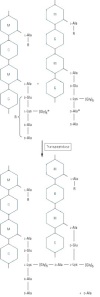
With such a wide range of activities against so many organisms one might wonder how they work.  Like all poisons, penicillins inhibit critical life processes in the species attacked. In this case, they inhibit bacterial growth by stopping the repair and replacement of their cell walls.  In affected strains, the cell walls are composed of long chains of six carbon sugars with amino acid side chains that weave the chains together like a net.  The long chains are made by polymerizing N-acetylglucosamine and N-acetlymuramic acid sugars into alternating units.  The side chains are made of repeating amino acid units, which differ per species, but for susceptible species they end with the combination D-alanyl-D-alanine amino acids.  B-Lactam antibiotics are natural analogues of these amino acids, which is to say they can fit into some of the same places as the natural bacterial amino acid piece can.  The antibiotics attach themselves to a special protein (the aptly named ‘penicillin binding protein’) that transports and tethers the D-alanyl-D-alanine amino acids to other chains in the cell wall, blocking its activity(S).  With these proteins blocked the cell wall can’t be replaced or repaired. Lysis results when the cell wall is unable to withstand the osmotic pressure upon it.

It is important to point out that these antibiotics are most active when bacteria are growing and synthesizing cell walls, which is fine for fast growing colonies, like the streptococci.  However mycobacteria, which are famous for causing tuberculosis and leprosy, have a dormant states where they don’t actively synthesize new cell wall components as well as impregnate them with certain lipids that impede the entry of penicillins.  In this context, an attack of this kind would be useless or at the very most a stop gap measure.  Since the colony isn’t completely killed off during treatment, this situation is favorable for the development of antibiotic resistances and the selection of hardy strains of the bacteria.  In a clinical setting these factors complicate issues, with treatments for tuberculosis taking upwards of a year or more to complete.

In a colony of 10^6 bacteria, the probability is that at least one will have resistance to a given antibiotic.  A typical mycobacterial colony will have something around 10^8 members, making the selection of strong, antibiotic resistant strains almost a certainty.  This problem is further compounded by a form of bacterial communication called horizontal gene transfer or plasmid exchange.  This is where a circular strand of DNA, typically encoding a gene producing a resistance, is absorbed by other bacteria allowing the technical means of resistance to spread. This is how bacteria such as the dreaded MRSA have developed and why infections from them are most common in hospitals. In the case of tuberculosis, multi-drug therapies utilizing non-B-lactam antibiotic combinations, like isonaizid-rifampin, can cure 95-98% of cases (S).  The probability that one will have a resistance against both antibiotics is very low and using two will usually kill the bacteria that have a selected immunity to one antibiotic alone.  The other 2-3% typically require specialized treatment and top of the line antibiotics, like moxifloxacin or rifabutin.  However, moxifloxacin resistant strains have been recently identified as a cause for concern (S).

Bacterial resistances usually present via four different routes: Reduced membrane permeability to the drug, production of an enzyme that deactivates the antibiotic (a B-lactamase, for example), a mutation in the PBP site where the antibiotic binds and finally, the creation of a means for drug removal (S).  With penicillins, B-Lactamase production is the most common form of resistance, some of which are highly specific to only one kind of drug (S).  B-lactamases attack the core structure of the penicillin molecule, breaking a critical carbon-nitrogen bond.  Mutations in penicillin binding proteins alter the shape of the binding site, making it harder for the B-lactam structure to fit.  In theory mutations could be great enough to block the penicillin from binding completely.  In practice, however, mutations that cause a PBP to be blocked entirely are often less useful functionally, because these will also block the D-alanyl-D-alanine amino acids they were originally intended for.  Small mutations are more useful to inhibit rather than block the penicillin and keep them from reaching a concentration necessary to inhibit bacterial growth. (S)  Gram negative bacteria usually develop resistances to antibiotic entry by changing the shape of receptors that transport the antibiotic across its membrane.  If the drug can’t pass through the membrane, it can’t attack the cell wall manufacturing machinery.  Another means that changes the membrane permeability is the creation of what is called an efflux pump.  These work to transport the antibiotics back across the membrane after entry.  These are of varied efficiencies, but always work to increase the amount of antibiotic required for efficacy.  Both mutation and changes in permeability are often paired with a B-Lactamase enzyme, making an effective, multi-front strategy for resistance.

Even though we have produced effective weapons against pathogenic bacteria, they have begun to fight back.  They have been largely successful in this regard, calling into question how long the usefulness of these drugs can be maintained.  This has spurred the demand for newer drugs to combat the potential ‘super-bugs’ on the horizon and limited use policies to maintain drug effectiveness in the future.  All in all, penicillin is generally considered non-toxic.  The worst adverse reactions are due to hypersensitivities.  Allergic reactions occur, however, this is a rarity occurring in less than 1% of people and is usually not life threatening (a rash, fever, etc).  Even less have a serious reaction, like anaphylactic shock (0.05% of patients).  In a worst-case scenario, though, desensitization to the drug can be used to effectively treat a given disease.

One thing is for sure is that we will continue to look in awe at the complexity life has to offer.  We hope that new attempts to cultivate and extract new antibiotics will be successful in the future.

{sources available upon request}

Read Full Post »

To live and function the human body needs energy, the majority of which is produced by the electron transport chain.  The end products from these enzyme catalyzed reactions are oxidized by the oxygen that you breathe and converted into water.  At the center of this complicated process is an incredible  protein that selectively transports oxygen to tissues in need and releases it on demand.  Hemoglobin is a protein carried by red blood cells that has selective affinity for iron, a metal critical to proper function of the body.  The importance of this protein is exemplified by what happens when it is broken.  Sickle Cell Anemia, for example, is caused by a mutation in the gene that codes for the protein.   In this case, the change of a single amino acid (glutamic acid to valine) damages its functionality such that the average life expectancies of people with this disease are only 42 years in males and 48 in women (S).  During this time, those afflicted also experience a host of quality of life maligning health problems, like chronic pain, increased risk of infection and heart disease and vaso-occlusive crises that occur when the misshapen blood cells block blood vessels.

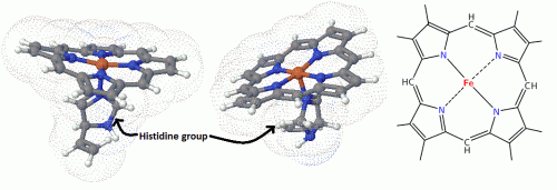
Considering the structure, hemoglobin is a large protein made of four polypeptide chains, sewn together into a tetramer.  There are 2 β chains and two α chains, each of which having a net-like porphyrin ring system.  In human red blood cells, an iron atom is placed in the center of the ring structure, creating a coordinated system called a heme group.  The iron atom is the star of the system and makes the binding of oxygen possible.  It is in the +2 state and has octahedral symmetry, allowing 6 ligands to bind.  The 4 nitrogen atoms of the ring bond equatorially holding the Fe+2 in place, while from the bottom, a histadine amino acid from the protein super structure locks the Fe+2 atom in place.  The open top position is reserved for O2 binding, but in its absence a molecule of water usually takes its place in normal circumstances.

3D images showing the positions of the porphyrin ring, iron and histidine in the heme group. Nitrogen atoms are in blue, iron in red and carbon in the usual grey-black. (3D images courtesy of http://www.3dchem.com By all means check out the structures for yourself using their tremendously useful java based web tool!)

This same system is common throughout other enzymes and respiratory systems.  In humans, Cytochrome C  of the electron transport chain has a similar structure, except that the 6th (top) ligand is a methionine group.  The protein in this case is designed to transport electrons rather than oxygen molecules.  Myoglobin, which is found in muscles and also used for O2 transport, is structurally different, but utilizes a single porphyrin ring system, rather than four.  Enzymes, such as catalase and peroxidase,  also contain Fe manipulating systems similar to the above.  In other kinds of organisms too, the porphyrin ring is utilized in a variety of situations and complexed to many different metals depending on the conditions and needs of the creature involved.  Photosynthetic plants, for example, utilize the same ring, but the metal is magnesium.  Further, some bacteria are also known to use copper as the porphyrin metal of choice.

To make full advantage of this heme system, the binding of O2 in red blood cells is facilitated by a buffer system.  The concentration of any of the components of it increase or decrease the binding affinity of O2 at the hemoglobin binding site.  Proton (H+), CO2, Cl-, and 2,3-Bisphosphoglycerate (BPG) concentrations all have a role in the binding and release of O2 from hemoglobin.  For example, in tissues where the pH (H+ concentration) is acidic and the partial pressure of CO2 is high, the binding affinity of O2 at the binding site will be lowered and will induce hemoglobin to release its contents.  In the lungs, however, the O2 concentration is high compared to that of CO2, facilitating O2 binding.  Normally, BPG is found in equal amounts to hemoglobin, but in situations where O2 is in short supply this balance is undone by the increased production of BPG.  With BPG, binding to one of the active sites reduces the O2 binding affinity by about 25 times, thus when BPG concentration is high it pushes for a release of O2 into tissues that need it.

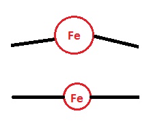
The larger Fe staggers the planar porphyrin ring, where as the smaller version fits better, contributing to stability.

Aside from outside effects promoting binding, there are effects coming essentially from the design of the system itself that have to be considered as well.  The question of why iron and not some other transition metal is of special relevance here.  Using iron as the central metal, in this case, yields benefits in terms of performance and binding specificity to oxygen.  Cobalt, for instance, is used in similar systems like those of vitamin B12’s corrin ring, but while both cobalt and iron contain d orbitals that could bind, only iron allows for the perfect balance between size and binding specificity in this system.  While unbound, the Fe+2 atom is just a little too large to fit into the normally planar porphyrin ring.  In this case, it is in a high spin state where the molecular orbitals are further away from the atom, giving it a larger size.  The high spin state is larger, because the Pauli exclusion principle prohibits the atom’s 3d electrons from getting too close to one another due to repulsions.  Thus, additional space is needed to house the electrons in orbitals further away.  However, when the Fe+2 binds to an oxygen molecule the electrons can be shuffled into orbitals that are closer to the atom.  Its orbital symmetry changes to a low spin symmetry and CLICK ! the Fe+2 shrinks just enough to fit snugly into the porphyrin ring system.

A silly, but useful comparison.

This part also contributes to a critically important finding: that the binding of O2 is cooperative, in that the binding of one O2 molecule will facilitate the binding of another until all four spaces are filled.  It has been experimentally determined that the binding energies (the Ka) are increasingly smaller for each molecule of O2 bound to hemoglobin.  This happens, in part, because of the protein’s structure.  When the O2 binds and the Fe+2 clicks into place, it pulls on the histadine residue below it, stretching the other protein superstructure bonds.  This pulls slightly on the other 3 Fe+2-histadine bonds.  Much like those dancing string toys, pulling on the string at the bottom causes the toy’s arms and legs to move in a concerted action, which is similar in a way to the physical reaction of the other binding sites.  The additional pulling on the other histadine residues facilitates binding of the other three, such that the binding energy is progressively reduced with each binding until all four slots are filled.

Other structural contributions also play a large role in binding specificity.  Above the plane of the porphyrin molecule lies another histadine residue that physically blocks the strongest and most effective bonding interactions from occurring.  Since the Fe+2 atom is locked into place, the best bonding interactions would come from ones that provide the most overlap of their molecular orbitals, which are those that are end-to-end.  However, with the histadine in the way, these are prevented from occurring.  This is a good thing, because strong covalent interactions in enzymatic reactions, like those seen in the binding of carbon monoxide (CO) for instance, are usually toxic and are difficult to break under normal conditions.  For head-to-head binding to CO, the interaction is estimated to approximately 1000 times as strong as those between O2, illustrating its toxic potential.  In this case, the CO-Fe binding is still strong, but not so much as to completely block removal.  People who have suffered CO inhalation are often given pure oxygen in an attempt to out pace CO binding and ensure that the person continues to have a supply of oxygen.  This hindered binding is also helpful in normal activity as well, since the binding symmetry to O2 is bent as well it further facilitates it’s release into tissues in need.

Yes, it’s about iron again, but it’s quite interesting stuff.

{sources available upon request}

Read Full Post »

Enterobactin

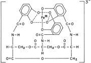
A little over 40 years ago it was discovered that bacteria utilize a class of metal chelators called siderophores to capture iron from their environments.  Iron is an important element for the growth and development of most kinds of bacteria.  It is used as a critical element in electron transport chains for energy production, the removal of dangerous reactive oxygen species and the functional element in the reactive sites of many important enzymes.  The finding was critically important to the field of medicine, as the iron scavenging activities of pathogenic bacteria are most often detrimental to the host.  Even though humans and other animals have developed sophisticated systems to sequester iron and make the body inhospitable to these bacteria, likewise they have developed a variety of means of out-competing their hosts for the valuable element.  Neisseria gonorrheae and N. meningitidis, for example, have evolved the ability to directly capture iron containing lactoferrin proteins from their human hosts, a testament to our continued co-evolution (1).  Still others have relied upon producing their own iron scavenging siderophores to fish out this valuable metal from fluid and protein sources.  Escherichia coli , for example, are known for producing enterobactin and have one of the most effective iron scavenging systems known, which at biological pH easily out competes endogenous defenses for iron.

Recently, it has been noted that these compounds are of critical importance to bacterial growth, especially in the ultra-low iron concentrations of bodily tissues.  Rather than tighten their belts in the toughest of times, some kinds of pathogenic bacteria turn on

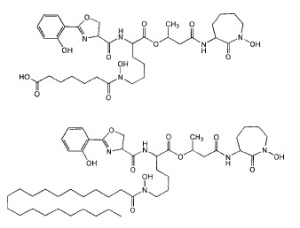
'Like dissolves like': different kinds of Mycobactin are used in different mediums. The long aliphatic chain endows fat solubility to the molecule, allowing it to shuttle through the waxy mycobacterial membrane easily.

these scavenging systems to forcibly acquire the iron needed for growth.  Mycobacterium tuberculosis produces several siderophores, called mycobactins, that are medium specific in their activities.  The two differ by a water soluble or fat soluble group attached to the siderophore skeleton.  Water-soluble mycobactin T is released into aqueous mediums, whereas fat soluble mycobactin T is designed to take pirated iron across the waxy mycobacterial cell membrane (2).  When iron is in good supply, however, production of these is unnecessary and is down-regulated to conserve valuable energy and resources.  Below the level of production of these siderophores, however, lies a regulated system of control.

Generally siderophore production is controlled by gram negative bacteria’s “Fur” and gram positive’s “DtxR” systems, which have  transcription elements that are bound by iron.  Thus, when intracellular iron concentrations fall below a threshold (approximately 10^-6 M for many pathogenic bacteria) the iron lynchpin holding back the transcription element is gone, activating the siderophore production system.  In 1999, researchers at the University of Queensland showed that Mycobacteria require the production of siderophores for growth in human tissues and macrophages (2).  They used genetic engineering techniques to remove the instructions for the enzyme MbtB, which is a critical component of mycobactin synthesis.  MtbB effects the final step in mycobactin synthesis, the coupling of a salicylic acid group to the siderophore’s finished structure.  After creating an MtbB knockout strain, its ability to produce siderophores was tested by an assay that measured the absorbance of the growth medium.  They used a colored dye compound that has a known affinity for iron in solution.  If a bacteria were to produce something that has a higher affinity for iron than the dye, this would be measured as a drop in absorbance, as the siderophore would compete with the dye for the iron.  They found that after comparing these bacteria against wild type strains that they were unable to produce siderophores and the deficient strains experienced retarded growth both in plate medium and in live macrophages (in vivo vs. in vitro tests).

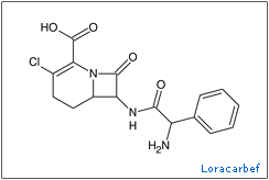
The understanding of how important these chemicals are for bacterial growth has led to renewed attempts to develop antibiotics against them.  In the late 80 and early 90’s the ‘trojan horse’ tactic of using siderophore-antibiotic conjugates and piggybacking them into a cell began to bear fruit with successes being discovered in a wide variety human diseases (3)(4)(5).  One group of scientists developed conjugates using the carbacephem antibiotic, loracarbef.  They found that the mixed catecholate-hydroximate siderophore they used gave the conjugate compound 2,000 times more potency than the parent drug alone (6).  Though resistances to this tactic still developed, these are thought to leave the bacteria at a disadvantage, because the mutations hinder the siderophore uptake system rather than attack the antibiotics themselves.  This limits access to a vital nutrient and leaves the mutants more susceptible to iron starvation and hindered growth.

Though the trojan horse tactic has yielded positive results, there are opportunities for even greater exploitation.  Techniques involving small molecules that attack that actual production of siderophores could provide another avenue to beneficial therapy for these diseases.  These drugs, much like statins that block the enzyme HMG-coA, would block a critical piece of the siderophore production pathway.  In the particular case of mycobactins, the final step in biosynthesis is the attachment of a salicylate group to the mycobactin skeleton.  Researchers at Cornell published results in 2005 showing that inhibitors of the enzymes that accomplish this task are potent inhibitors of M. tuberculosis and Yersinia pestis (7).  They synthesized a compound, SAL-AMS, that closely resembled a reaction intermediate and measured its effect upon the growth of bacterial cultures in mediums of low iron concentration.  They found that it successfully inhibited the enzyme and drastically reduced bacterial growth in the cultures.  It was shown to have an IC50(*) of 2.2 ± 0.3 μM for M. tuberculosis and about 51.2 ± 4.7 μM for Y. pestis in an iron limited medium.  Though in mediums with high concentration of iron the chemical was ineffective against Y. pestis, but it was found that the chemical might have unknown inhibitory properties against M. tuberculosis, as:

“Salicyl-AMS (tested at up to 8 X IC 50) was not active against Y. pestis in iron-supplemented medium, in which siderophore production is not required for growth.  Under these conditions, salicyl-AMS (tested at up to 180 x IC50) did inhibit M. tuberculosis growth, albeit with an 18-fold increase in IC50 (39.9 ± 7.6 μM).  This suggests that, in addition to blocking siderophore biosynthesis, salicyl-AMS may also inhibit M. tuberculosis growth by other mechanisms.”

Furthermore, researchers at the university of Minnesota developed similar nucleoside inhibitors of MbtA, one of which ” rivals the first-line antitubercular isoniazid” in activity against the bacteria (8).  This tactic follows the same reasoning as SAL-AMP above, as the nucleoside inhibitors attack the same pathway to inhibit siderophore end production.  The research was centered around making logical functional modifications to known structures of inhibitors and choosing the most effective ones for further testing.

Comparison of the pathway intermediate, the Cornell inhibitor and one of the Minnesota nucleoside inhibitors.

These findings are coming just in the nick of time it seems, as certain drug resistant strains of M. tuberculosis have become big news recently.  Extensively drug resistant tuberculosis is a kind of tuberculosis that is resistant to at least two of the top line drugs used to normally treat it (typically isoniazid and/or rifampicin) and a member of the quinolone antibiotics (ciprofloxacin).  Tuberculosis is generally a challenge to treat in the first place, with treatments typically taking up to a year or more to complete.  The loss of the first line drugs and reliance upon second line increases the risk of side effects and patient noncompliance to the already long course of therapy.    This can further complicate the issue, as it could lead to the obsolescence of the few active  drugs used to treat the disease, because resistances to one drug are usually useful against the whole family of drugs.

β-lactamases typically attack the carbonyl in the β-lactam structure, destroying the ring. Nafcillin has a large group that hinders these enzymes from getting too close.

An example of this can be seen in bacteria that produce β-lactamases, as these strains are often cross-resistant to all unprotected β-lactam antibiotics.  Some  penicillins have been designed with bulky groups attached to the skeleton in an attempt to hinder these enzymes.  Nafcillin, with its large 2-ethoxy-1-naphthoyl group, is very effective at blocking these enzymes for the most part.  However, even this tactic has its limits as methicillin resistant Staphylococcus aureus (MRSA) and oxacillin resistant Staphylococcus aureus (ORSA), both have developed resistances against these drugs such that, “From 1999 through 2005, the estimated number of S. aureus–related hospitalizations increased 62%, from 294,570 to 477,927 (9),” and “MRSA accounts for an estimated 12% of all nosocomial bacteremias, 28% of surgical wound infections, and 21% of nosocomial skin infections.  Infections secondary to MRSA result in excess costs of approximately $4,000 per patient per hospitalization compared with patients infected with methicillin-susceptible S aureus (MSSA)”(10)(11).

Since the discovery of penicillin, Alexander Fleming hypothesized that bacteria would develop resistances to the antibiotics used to treat them.  Now, more than ever, the development of new antibiotics must be pursued.  Human defenses have always provoked a counter-response from our pathogens, the most successful tactic selected out.  Much like the pathogens that infect us, we must ‘evolve’ a newer understanding of bacterial biology, which will offer us a foothold to better, more effective treatments.

——

(1) Genetics and Molecular Biology of Siderophore-Mediated Iron Transport in Bacteria

JORGE H. CROSA

(2) The salicylate-derived mycobactin siderophores of Mycobacterium tuberculosis are essential for growth in macrophages

James J. De Voss, Kerry Rutter, Benjamin G. Schroeder, Hua Su, YaQi Zhu, and Clifton E. Barry III

(3) Design, Synthesis, and Study of a Mycobactin−Artemisinin Conjugate That Has Selective and Potent Activity against Tuberculosis and Malaria

Marvin J. Miller, Andrew J. Walz, Helen Zhu, Chunrui Wu,Garrett Moraski, Ute MÖllmann, Esther M. Tristani, Alvin L. Crumbliss, Michael T. Ferdig, Lisa Checkley, Rachel L. Edwards, and Helena I. Boshoff

(4) Species Selectivity of New Siderophore-Drug Conjugates That Use Specific Iron Uptake for Entry into Bacteria

M. S. DIARRA, M. C. LAVOIE, M. JACQUES, I. DARWISH, E. K. DOLENCE, J. A. DOLENCE, A. GHOSH, M. GHOSH, M. J. MILLER,    AND F. MALOUIN

(5) Siderophore-Based Iron Acquisition and Pathogen Control

Miethke M, Marahiel MA.

(6) Iron Transport-Mediated Antibacterial Activity of and Development of Resistance to Hydroxamate and Catechol Siderophore-   Carbacephalosporin Conjugates

ALBERT A. MINNICK, JULIA A. McKEE, E. KURT DOLENCE, AND MARVIN J. MILLER

(7) Small-molecule inhibition of siderophore biosynthesis in Mycobacterium tuberculosis and Yersinia pestis

Julian A Ferreras, Jae-Sang Ryu, Federico Di Lello, Derek S Tan & Luis E N Quadri

(8) Antitubercular Nucleosides That Inhibit Siderophore Biosynthesis: SAR of the Glycosyl Domain

Ravindranadh V. Somu, Daniel J. Wilson, Eric M. Bennett, Helena I. Boshoff, Laura Celia, Brian J. Beck, Clifton E. Barry, III, and Courtney C. Aldrich

(9) Hospitalizations and Deaths Caused by Methicillin-Resistant Staphylococcus aureus, United States, 1999–2005

Eili Klein, David L. Smith, and Ramanan Laxminarayan

(10) Baquero F. Gram-positive resistance: Challenge for the development of new antibiotics. J Antimicrob Chemother. 1997;39(suppl A):1–6.

(11) Kopp BJ, Nix DE, Armstrong EP. Clinical and economic analysis of methicillin-susceptible and -resistant Staphylococcus aureus infections. Ann Pharmacother. 2004;38:1377–1382.

(*) The IC 50 is the half maximum inhibitory concentration of antibiotics, a typical measure of effectiveness of the drug.

Read Full Post »

Polyethers are made by the anionic polymerization of an epoxide and a strong base like an alkoxide ion. This reaction polymerizes the two compounds and creates repeating ether (R-O-R) links between them making long chain compounds from 300 g/mol to 10,000,000 g/mol. This kind of reaction is ended when an alcohol like methanol deactivates the end chain anion by donating a hydrogen atom. As for physical properties, they have a high solubility in water because of their ability to make hydrogen bonds. Industrially they are marketed under many names, but a carbowax (varied types) seems to be the most popular name.

These chemicals have a surprising amount of uses as surfactants in industry, including foods (filler: probably McD’s), cosmetics and pharmaceutics; in biomedicine, as dispersing agents (timed release medicines), solvents, ointment (the lube, dude: KY) and suppository bases (GoLYTELY, GlycoLax), vehicles (car waxes), new types of body armor (liquid sheer thinking fluid body armors), cloth and fabrics (think spandex), tablet excipients (inactive ingredients in vitamins medicines). They have a low toxicity (1) and are finding new use in the repair of damaged nerve cells in animal trials (2), as well as use as a colo-rectal cancer preventative.

Unfortunately, the human trials are too late for Christopher Reeve and others who suffer a similar fate, but perhaps they will happen sooner than we think and hopefully (if we’re lucky the research is applicable to humans) many will reap the rewards of this research. Trials have been conducted with guinea pigs, dogs and such with fairly encouraging results (2), but the effect on human damage is unknown. One thing is certain: its definitely not an ordinary laxative.

1. Toxicology

http://www.mindfully.org/Plastic/Pol…ycols-PEGs.htm

2. Animal tests:

http://www3.interscience.wiley.com/c…TRY=1&SRETRY=0
http://www3.interscience.wiley.com/c…06760/ABSTRACT
though not applicable to humans, this link talks about its use as a colorectal cancer preventative:
http://www.inra.fr/reseau-nacre/sci-…t/indexan.html

3. Nerve guides help regeneration

http://www3.interscience.wiley.com/c…97042/ABSTRACT
http://www3.interscience.wiley.com/c…32586/ABSTRACT

4. general interest news

http://seattlepi.nwsource.com/health…_spinal04.html

Read Full Post »

As I was running yesterday a thought occurred to me as the steady, percussive hum of wings buzzed by me several times: one giant Asian hornet (an osuzumebachi) is startling, but several dozen is pretty scary. Apparently, I had run too close to a nest and had to hot foot it out; I was lucky.

These things are pretty amazing creatures, with an average body length of 2 inches and a wing span of 3, you will stand up and take notice when they fly past. As with many bee and hornet stings single stings can be deadly as a result of anaphylactic shock, but this is fortunately a rare occurrence. Nonetheless, the hornets are nothing to take lightly as the venom contains an interesting mix of nastiness, as well as a barbless stinger, which it can use repeatedly unlike regular honey bees. The venom contains neurotransmitters like serotonin and acetylcholine, which is responsible for pain transmission, a neurotoxin called mandaratoxin, tissue dissolving enzymes such as phospholipase-B, a mast cell degranulating peptide called mastoparan and a pheromone that instigates other hornets to swarm and attack. Aside from pain, the stings usually leave a dime-sized scar not unlike those of certain poisonous spiders in the southwestern US.

The hornets terrorize smaller, weaker prey like praying mantii and Japanese honey bees. The bee’s stingers are too small to penetrate the hornet’s tough, armored exoskeleton and thus have little defense from a full on attack. They reputably work with ruthless efficiency, a single hornet able to kill as many as 40 bees per minute and a few hornets only a few hours to slaughter a 30,000 bee colony. Interestingly, the bees have a small defense against them in their greater tolerance for heat. Typically, the bees will entice a scout inside the hive, where they will attempt to smother him in a bull rush. Once he is covered, the bees will vibrate their flight muscles increasing the heat in the area to around 47C. The hornets can’t survive past 45C, preventing the scout from calling reinforcements.

Link: http://www.youtube.com/watch?v=JDSf3Kshq1M
Link: http://www.youtube.com/watch?v=hpcHH1EpTZM

Even though, these may seem like kings of the forest, human initiative has once again found ways to make use of nature. The Japanese love energy drinks, one of the more famous products, VAAM is of particular note. As advertising shows it has been the favorite of many a marathon runner and endurance athlete. Yet a little less advertised fact is that it’s a chemical copy of the stomach contents of these hornets. More specifically, it is a copy of the secretions of the colony’s larvae, which the adults feed upon as they lack the ability to digest raw protein themselves. How’s that for wild? This mixture of predigested amino acids and sugars supposedly allows them to fly distances of 60 miles per day at rates of 25-40 miles per hour.

Sounds perfect for runners, now lets have a toast!

As gross as that sounds many professional athletes here swear by its supposed benefits and from personal experience it tastes pretty good. The question of the day, though, is one of efficacy: Does it work? Dr. Takashi Abe of the Institute of Physical and Chemical Research in Japan seems to think so. “VAAM works by helping the body burn the energy it stores more efficiently”…”[It] expedites the metabolism of fat and promotes better hydration,” said Dr. Abe. Animal studies conducted by Abe concluded that mice that were fed VAAM could swim twice as long as those that were fed only water and 25% longer than those fed casein, a protein found in milk. Their blood also contained fewer fatty acids than those of the other groups, an indication of how efficiently the mice were burning their fat reserves. Details are sketchy as to how it might actually accomplish these feats. One possible route may be through the branched chain amino acids in it, which increase the effect of insulin in the body. The product contains a mixture of leucine, isoleucine and valine, of which leucine is considered the most effective at this. Leucine reportedly “increases insulin output by 221%,” if taken with carbohydrates after exercise.

If you’re thinking: “At last the perfect supplement to my admittedly weak dietary habits, that will propel me to epic heights of fame and fortune,” then you’re most likely going to be disappointed (mostly in yourself). I’ll save you the trouble and clue you in to something important: the supplement industry is mostly a joke played on the lazy for the fun of a few bored chemists. Though there might be a slight benefit to some supplementation, there is no substitute for hard work, but we delight in trying to make you think there is. (Next year’s big craze will be whale sperm marketed under the name SPEaRM—->.) On the other hand, if you’re looking to try something that tastes good to replenish your vital fluids during or after a workout, then this might be up your alley.

Read Full Post »

In 1913, a Swiss chemist named Alfred Werner was awarded the Noble Prize in Chemistry for his work on what would be called

Vanadium rainbow.

coordination chemistry, which would lead to a new understanding of how chemicals bond together. Coordination theory describes the nature of bonding in transitional metals and the formation of complexes, which at the time seemed to follow bizarre and unpredictable patterns. Atoms in groups 1-7A followed somewhat predictable patterns in their bonding as shown by classical experiments. For example, atoms in group I took on +1 charges and bonded once to other more negatively charged atoms like the group 7A halogens, but it remained a mystery how the transition metals bonded and why they had so many oxidation states.  Vanadium, for example, produces a wonderful rainbow of oxidation states when potassium permanganate is added to a Vanadium II solution.  Over time it separates into different states: V +2 is violet, V +3 is green, VO +2 is blue, VO2 +1 is pale yellow, MnO2 is brown and MnO4 –1 is pink.  In Werner’s time, the shape of salts like (CoCl3 * 6 NH3) were still undetermined and throughout much of the 1800’s one popular theory emerged: chain theory. It was supported by some of the most powerful chemist-sorcerers at the time, including Werner’s chief rival: S.M. Jorgensen.

Jorgensen believed that the ligands in compounds like (CoCl3 * 4 NH3) were arranged in chains, that is, bonded to each other in some fashion. The main point being that the atoms would follow known valence rules at the time, especially Kekule’s principle, which abstracted the number of times a compound could bond from known chemical reactions. Though useful, it ran into problems when trying to describe why atoms with larger electronic configurations bonded in so many different arrangements. Transitional metals in particular confounded these rule sets.

Werner, however, proposed a different theory that relied on the concept that cobalt (in the above compound) could have more than the three bonds predicted by Kekule’s theory and that the ligands would be centered around cobalt in an octahedrally arrangement, rather than in chains. According to his theory, a compound like the above due to its structure would have two possible conformations: a cis isomer (with chlorine atoms on adjacent vertices) and a more stability favored trans isomer (with the chlorine atoms on opposite side of one another). Interestingly, the two are identifiable by their color with the trans compound being green and cis being a delightful purple color

Naturally, this caused controversy amongst chemists and the debate began. At the time only the structurally favored green trans compound had been synthesized, while the more difficult cis compound was thought to be non-existent.  Cis compounds are generally less stable and in this case it is due to repulsions between the electronegative chlorine atoms positioned close to each other.  Whenever Werner published results that seemingly confirmed his theory, Jorgensen was there to propose a counter theory in favor of the more popular chain theory.   Chain theory had strength in the fact that there are many possibilities in the way ligands can be arranged in that manner.  Eventually, Werner was able to prove his case conclusively through a variety of methods like optical resolution of the compounds and electrical conductivity measurements. The capstone, as the story goes, was his synthesis of the elusive purple cis isomer of [Co (NH3)4 Cl3] and sending a sample through the mail to Jorgensen.  The flurry of high fives and  chest bumps went unabated for three months afterward and was actually seismically measured in Sweden.


Werner used this clever method to synthesize his purple cis isomer. By adding HCL at 0C, carbon dioxide is released and chlorine atoms in solution replace the oxygen atoms lost. (Note: picture does not show positive charge on Cobalt atom)

Transition metal like many of the blue colored above are used in a variety of reactions ranging from biological (Zn, Co, Cu, etc) to industrial (Os, V, Pb, Pt, etc).

Read Full Post »

With over half a billion deaths attributed annually, Malaria is one of the world’s worst infectious diseases. The WHO, spurred on by previous success with small pox, has long had the dream of eradicating this disease, but has had to deal with limited success as the mosquitoes have developed resistances to the insecticides traditionally used against this primary vector. Currently, the Global Malaria Action Plan (GMAP) has had ambitious plans “to spray 172 million homes and distribute 730 million insecticide impregnated nets” by 2010, but there is a hidden danger to the plan: natural selection. More and more we are finding that the traditional insecticides (DDT, Pyrethrins, etc) are becoming less able to control populations of Anopheles mosquitoes, because the selective pressure on populations is already powerful even without intervention.

The long term benefits of using ‘quick kill’ insecticides are limited, because they add to the selective pressure already existent in the normal breeding cycle, thus quickly creating populations that are resistant to these traditional methods. The problem is one of diminishing returns. Consider the impracticality of perpetually designing an endless chain of active, weakly human toxic insecticides ad infinitum. Insecticides are toxic chemicals by their nature and very few have been found to date that have the necessarily low human toxicity and insect killing capacity. This paper suggests that using new or old pesticides in new ways might limit or all together remove this selective pressure, while at the same time accomplish the stated objective of controlling malaria.

The Big Idea: How They Work:

Malaria is caused by a eukaryotic protest called Plasmodium falciparum, which is transmitted by the bites of Anopheles mosquitoes. Their life cycle is an important part of the problem. After eating, the females produce and deposit a batch of eggs in water, which in total takes 2-4 days to complete. Even without insecticides exacerbating the problem, the egg mortality rate is around 20-40% per batch. With such a turnover rate, breeding selection will always be a strong factor in mosquito populations. At some point, the mosquitoes encounter and become infected with the malarial parasites. These generally go through an incubation period of 10-14 days before becoming active in mosquito salivary glands. This is ironic considering that most mosquitoes will not live long enough to become infectious and spread the disease. Since they only spread the disease at late stages, there is a window of opportunity to curb not only the selective pressure that encourages resistances, but also dramatically control the malaria transmitting mosquitoes.

Late Life Acting insecticides (LLA) have been proposed that disproportionately kill only the older mosquitoes. A major benefit being that selective resistances would spread much slower, than as with the use of current insecticides, while at the same time cull the infective mosquitoes. Conventional insecticides reduce the mosquito reproductive success by about 85% at first, but the LLA’s, which would allow for a normal reproductive cycle would target only older mosquitoes. Selectively targeting only the older mosquitoes relieves this selective pressure. If only the older insects are killed, it allows the younger ones to proliferate and spread their genes giving little selective benefit for having a resistance to the insecticides. Furthermore, the authors point out that:

The strength of selection declines with age. Beneficial genes that act late in life can fail to spread if they are associated with fitness costs earlier in life.

Though LLA insecticides would leave a large population of mosquitoes, the key point is this is meant to be disease control, rather than insect control.

Several methods have been proposed. First, cumulative exposure to ordinarily sub-lethal doses of pesticides. The idea is that the low doses build up over time killing only the older mosquitoes before they transmit the disease. Second, micro-encapsulation techniques designed to release over a long period of time. (Think about your Imodium 24 hour extended release, except with horrible poison!!) Third, chemicals that negatively affect detoxification pathways in mosquitoes. This exploits the fact that as mosquitoes age they become less able to detoxify chemicals. Fourth, compounds or dosages of pesticides that take advantage of an infected mosquitoes weakened state (malarial parasites negatively affect them as well). By using dosages, which are lower than recommended (especially those which are non-lethal for most healthy mosquitoes), it may be possible to target only those older mosquitoes with a weakened composition. Fifth, fungal bio-pesticides, which are active against mosquitoes, killing them 7-14 days after contact. The final possibility mentioned is using Wolbachia bacteria or a densovirus to control the older populations. Wolbachia are inherited bacteria that infect many kinds of insects, in particular affecting the reproduction system of their hosts and in some species causing parthogenesis (reproducing only one sex). Densovirii are virii that are thought to only infect insects. Both of these could be applied in much the same manner as the above methods, either building up over time or causing a metabolic pitfall as they age, shortening their life spans to die before the malarial parasites become active.

Source:

Evolution Proof Insecticides for Malaria Control

Penelope A. Lynch, Andrew F. Read and Matthew B. Thomas

Read Full Post »

First and foremost, cancer is a disease of broken genes. For as long as medicine has existed, cancer has defied our attempts to cure it. It is estimated that one in 3 people will experience cancer in their life time(1). More so, “greater than 60 years ago there were no treatments for children with leukemia that could cause remission of the disease, but today this is possible in about 96% of cases!”(2) Though we have made leaps in our understanding and treatment of cancer, the golden fleece: freedom from this oldest plague of man has long eluded us. Perhaps some day we will achieve this ultimate prize, but it seems for the time being we must be satisfied with our current progress. Here’s a few reasons why the Tigers still stalk us in the halls when Sally Kimball is away:

1. Biological Immortality and unlimited growth:

To keep cells fit and in good working order, ordinary cells are limited by how many times they can divide. This is known as the Hayflick limit. A cell’s DNA has a non-coding region at the end of the strand called the telomere, which can be likened to a docking station for the DNA replication machinery. Every time the strands are copied this machinery takes a small piece of the telomere leaving behind a smaller and smaller piece. Once the telomere runs out, the cell enters senescence, stops cell division and dies. Somatic cells have about 50 divisions in them before this happens.

In cancer cells, an enzyme called telomerase actively replaces these lost pieces of telomeres, allowing them to divide beyond the Hayflick limit and evade the natural cell death program. Normally, this limitation keeps a cell line’s genetic package healthy and mutations from accumulating. However, for cancer cells this is not a problem, as the mutations sometimes serve to provide a survival advantage over normal cells. The tumor suppressing genes Rb1 and TP53, for example, are known to have roles in controlling cell growth, but once they are damaged or turned off they allow a cell to break free of the systemic growth of normal cells.

Retinoblastoma protein (Rb), the protein produced by the Rb gene, does its work by prohibiting a cell with damaged DNA from replicating. When this situation occurs, it binds and blocks the transcriptional machinery in cells, keeping it from copying the genome and consequently, proceeding into the next stage of cell division. Many cancer cells have mutations to this gene. In fact, the protein produced by this gene was discovered in a kind of eye cancer called retinoblastoma. This should not be misconstrued as only affecting the eyes, however, as it is a significant regulatory protein in almost every cell in the body.

P-53’s main job is to protect the genome from mutation. It acts as a watchdog right before cell division, making sure that the DNA has been faithfully reproduced. If there is DNA damage, then P-53 can stop the cell cycle and activate DNA repair proteins that can hopefully fix the damage. If the damage is too extensive to repair, then it can also signal apoptosis, the cell’s death program. Without this protein functioning properly, the risk of developing cancer is increased greatly. More so, “greater than 50% of all human cancers contain a deletion or mutation of the TP53 gene” (3).

Mutations of these genes generally allow a cell unrestrained, unlimited growth and are a cause of the massive growth of cells, known as a tumor. In this situation, cancerous cells have an advantage over normal cells, in that they have easier access to nutrient resources from the surrounding tissues.  They will grow in size until they are limited only by the availability of resources. An interesting observation is that the inner portion of many tumors is lower in oxygen (hypoxic), than the outer portions. This is a direct result of Fick’s law of diffusion, which describes the flow of nutrients and oxygen from blood vessels into tissues. It shows that as a tumor grows increasingly larger, it becomes harder to provide cells further away from blood vessels nutrition. This is a natural limitation on cell growth and body structure, but tumor cells have a way to bend this rule, which is described in the next section.

2. Decreased dependence on natural growth factors:

Cancer cells are like ‘rebels without a cause’, the James Deans of human cells. Normally, the signals for growth are usually sent by the endocrine/paracrine system. Platelet derived growth factor, epidermal growth factor and insulin-like growth factor are just a few regulatory chemicals that trigger cell division and cell proliferation. The collective mass of these signals make up a system that tells cells when and under what circumstances to begin division. To have a body and the organizational benefits that it brings, individual cells must perform the tasks they’ve been assigned in the developmental process. Cancer cells, however, produce their own growth factors, often self signal and/or develop a means to receive more signaling. Since they require no outside signals for growth, they are not limited by this systematic control. They effectively work outside the system to effect their own self centered proliferation, at the expense of the body supporting them.

One example of this is an activity called angiogenesis, the production of new capillaries and blood vessels from main branches. Normally, fetal tissue relies on the function of vascular endothelial growth factor (VEGF) to produce new capillaries in its rapidly growing tissues. More capillaries means that a greater volume of nutrients and oxygen can diffuse to important tissues. In adults this process is usually indicative of cancerous activity, with wound healing and the female reproductive cycle being the major exceptions. Tumors produce VEGF to get around the limitations posed by Fick’s law of diffusion. Tumor growth is limited to the short distance nutrients and oxygen can diffuse through tissues. Without the production of new blood vessels the mass of tumor cells would slowly starve.

VEGF has been found in almost every kind of human tumor and is at peak concentration around newly formed blood vessels and the hypoxic, inner regions of the tumors. Research has shown that by blocking VEGF by binding it to monoclonal antibodies, one can suppress tumor growth in mice(4). Other efforts are currently targeting VEGF receptors that are specific to kinds of tumors. Inhibition of a VEGF receptor called FLK1 has been similarly shown to reduce growth of tumors of certain cancer cell lines such as fibrosarcoma cell line HT-1080(5).

Another method cancer cells use to circumvent normal regulation involves the over expression of epidermal growth factor receptors (6). Normally, epidermal growth factor signals limited cell proliferation and its receptor is usually expressed in epithelial cells. However, cancer cells over-express it allowing for greater than normal signaling and as a result, increased proliferation. This over-expression is usually indicative of a poor prognosis, a situation occurring in many advanced stage cancers. In certain types of breast cancer, which comprise a little more than 20% of the total number of cases, the EGF receptor uses a protein called ‘HER’ to pass on the growth signal inside the cell. The bio-engineering giant Genentech recently made headlines for passing its drug herceptin through clinical trials. This is a monoclonal antibody that specifically targets HER proteins. Since cancer cells that over express EGF receptors have a greater preponderance of this protein, they will be targeted for destruction by the immune system specifically over ordinary cells.

Recent drug advances have allowed for the selective targeting of these receptors, effectively blocking this excessive, cancerous signaling. Specifically, several new tactics have been devised to target these receptors and block their activation. Recent successes with monoclonal antibodies, selective toxins and small molecules that block receptor signaling activity have made it into general therapy. Monoclonal antibodies are specifically targeted to these receptors to bind and block them from being activated. Toxins can take a variety of roles to block receptor activity, whether as allosteric inhibitors or by covalently binding to the receptor’s active site. Essentially, the end result is the same with these two kinds of weapons. With small molecule drugs, the tactic is usually to inhibit signal transduction from the receptor. The drug erlotnib is an example of this, in which it blocks a critical signaling component so signals can’t be sent from the receptor. Erlotnib is from a class of drugs called tyrosine kinase inhibitors, that have found great usefulness as selective inhibitors of these kind of over-expressed tumor receptors.

3. Loss of anchorage in tissues and cell adhesion:

Normally, cells will undergo apoptosis once they become separated from their host tissue. It’s tricky to grow normal cells outside of their home tissues, because they communicate with neighboring cells and require paracrine interaction for normal function. However, cancerous cells don’t require these regulatory signals and can be grown in solution or on agar medium. Normal cells are usually held in place by a matrix of proteins and structural elements, as well as being linked directly to neighboring cells by physical junctions. These structural components often give important regulatory information to a cell. The point where this breaks down is the beginning of what is called metastasis, the ability of a cancer cell to travel to other tissues, invade them and start a tumor there.

Tumor cells express a set of metal containing enzymes called matrix metalloproteinases (MMPs). These are responsible for the matrix degradation seen in metastatic cancers. They chop away at the extracellular matrix holding them in place, eventually freeing the cells to do damage elsewhere. The expression of these proteins is considered indicative of progression to metastasis, because ordinary adult cells do not express them. High levels of matrilysin (MMP-7), for example, is a known indicator of prostate cancer. Additionally, MMPs are known for attacking cell to cell adhesion proteins like E-cadherins, β-catenin and α-catenin. MMP-3, also known as stromelysin, is known for cutting apart E-cadherin cell junctions. The loss of their function is a known cause of tumorigenicity and cancer cell invasiveness.

Once a tumor cell escapes it has two routes it can metastasize through: blood or lymphatic. The tumors often have patterns of invasion that are indicative of their tissue origin. For example, tumors of the head and neck are often spread through regional lymph nodes (7). Tumor cells manufacture special tools they use to attach to new host tissues called invadopodia. These are similar in a way to bacterial pilli in that they contain various proteases and adhesive proteins that aid in attachment to the new cells and help the cell cross barriers.

The invadopodia bind to membrane components such as laminin, fibronectin, type IV collagen, and proteoglycans. Normally, these interact with receptor regulatory proteins called integrins that send regulatory signals to the cell. Cancer cells often change the binding preferences of their integrin receptor subunits to match those of degraded extracellular matrix proteins. The new types are thought to match the pieces damaged by MMP degradation and are often associated with invasive, metastatic cells (8). Binding to these pieces functions as a discovery signal that the cell has found a place that is susceptible to invasion. The fact that cancer cell integrin expression has changed in a distinct way, might also reveal a way to specifically target these cancerous cells. This could lead to therapeutic advances targeting the most aggressive, late stage types of cancers.

4. Loss of sensitivity to apoptotic stimuli:

Apoptosis is a form of cell death that is essential for tissue remodeling during embryogenesis and maintaining the number of cells in adult life. It functions to cull damaged, nonviable and potentially dangerous cells by acting as a failsafe that kills a cell when it has acquired too many mutations or unusual activities. It functions through cellular signaling pathways to block cell mitochondrial function, which eventually leads to the degradation of critical cell components and then ultimately, death.

The apoptotic program can be divided into three phases: initiation phase, decision/effector phase and the degradation/execution phase. The initiation phase typically is a response phase to outside stimuli, like death receptor ligands, or to inside stimuli like DNA damage. The decision/effector phase works to clarify the signal and open the door for action. Changes that occur in mitochondrial membrane permeability signal the end: the release of an key respiratory protein called cytochrome C into the cytoplasm. The degradation/ execution phase sees the activation of proteases and nucleases, which degrade proteins and nucleic acids, respectively. The key target is the mitochondrion, as well as other important cell machinery. As this is the main power source for the cell, this is typically fatal for it.

Apoptosis acts through signal transduction pathways controlled by receptors of the Fas cluster (CD95), tumor necrosis factor receptor 1 (TNRF1) and death receptors 3, 4 and 5 (DR 3, 4, 5). Each of these kind of receptors contains a special amino acid sequence called the “death domain” that functions as a specific binding site for special death signaling proteins. These proteins pass the baton on to other proteins in the chain, much like a relay. Eventually they activate the main downstream effectors, the actual ‘doers’ of the hard work, the caspases. Caspases are a family of cysteine proteases that are responsible for much of the cellular degradation mentioned earlier.

The point of no return in this deadly relay is the release of cytochrome C, which is controlled by a set of proteins in the mitochondrial membrane that regulate its release. The first of these was identified in a cancer called B-cell lymphoma and was aptly named the B-cell lymphoma-2 (Bcl-2) protein. This particular protein is a negative regulator of apoptotic signals. This group of regulatory proteins contains both positive (Bax, Bak, Bik, Bid) and negative regulators (Bcl-2, Bcl-x) of these signals. Positive regulators encourage apoptosis, whereas negative regulators discourage it. The key factor determining cytochrome C release is the relative ratio of positive and negative signals.

Cancer cells evade apoptosis by overexpression of Bcl-2. The Bcl-2 gene is usually moved to a different, more active location in the chromosome (typically the IgH promoter, a highly active portion). With more of this protein floating around, the pro-apoptotic signals are drowned out and sensitivity to them is significantly reduced. Lastly, cancer cells suppress apoptotic receptors by mutations that affect binding and proper function of the pathway. Slight mutations to the receptors or relay proteins can drastically affect the ability of the apoptotic signal to reach the critical stage.

5. Genetic instability:

Genetic instability is considered a major causative problem in cancers and one that we have few effective weapons against. Our battle strategy is reactive and revolves around destroying the cells that display these characteristics when they become a noticeable problem, which is often the time with the least efficacy in treatment. A truly proactive strategy will require advances in technology and bioengineering that will enable us to manipulate our genetic code, to edit out or silence problematic parts and correct mutations when they occur. The promise of gene therapy holds the hopes of many to pick up and wear that mantle, but these experiments are a ways off from useable therapies. Today though, like any good general, we make do with the tools we have on hand.

Cancer cells are often distinguished from normal cells by the loss or gain of a specific chromosome, or even the accumulation of an entire extra set of chromosomes. Extra chromosomes can provide cancer cells with extra copies of growth promoting genes. They can utilize these extra copies to amplify ordinary levels of signaling, causing the increased growth response and proliferation seen in tumors. Mutation is common in human tumors where changes at the sequence level can affect growth controlling genes, DNA repair or decreased fidelity during replication. Furthermore, it has long been known that misreplicated DNA can provide a causative explanation for some inherited cancer prone syndromes.

Translocations of genetic material from different chromosomes can lead to the abnormal gene expression seen in cancer. Chromosomal rearrangements, for example, are known to cause cancers like chronic myelogenous leukemia (CML) and Burkitt’s lymphoma. CML is caused by an abnormal chromosome, called the Philadelphia chromosome (named after two scientists from Philly). In CML, two unrelated genes from chromosomes 9 and 22 switch places and parts of the genes are spliced together. This causes a new protein called “BCR/abl” to be produced that remains constantly active, driving cell division by activating cell cycle control proteins continuously and inhibiting DNA repair responses.

Fortunately, since CML is caused by a single protein, it represents a good target for drug based therapeutics. New tyrosine kinase inhibitors, like imatinib, are used to block BCR/abl’s activity and has become the standard treatment for the disease over previous less specific antimetabolite chemotherapies and bone marrow transplants. This drug represents a different level of complexity in the treatment of the disease. An intimate molecular understanding of the disease, lead to the search for drugs that could effectively inhibit the protein’s activity. Drug designers searched protein libraries, through thousands of possible candidates to find the best ones, eventually coming up with imatinib.

Imatinib is representative of a new kind of drug design, where a drug is designed from known specifications of the biological machinery. Specific understanding of the processes involved will ideally improve drug activity and binding specificity and hopefully open the door to more effective therapies. Even though it can not cure this disease, it makes management of the disease possible and improves the lives of patients over that of previous chemotherapy based treatments.

6. The Loss of Cell Cycle Control: Until next time…

Some sources:

(1)http://en.wikipedia.org/wiki/Cancer (In epidemiology section.)

(2) Gale encyclopedia of cancer, pg. 11 intro, Helen A. Pass, M.D., F.A.C.S.

(3) http://en.wikipedia.org/wiki/P53

(4) ask me later, lost it…

(5)B. Millauer, M. P. Longhi, K. H. Plate, L. K. Shawver, W. Risau, A. Ullrich, and L. M. Strawn, Cancer Res., 56, 1615-1620 (1996).

(6) 60. N. Ferrara, and W. J. Henzel, Biochem. Biophys. Res. Commun., 161,851-858 (1989).

(7) Burger’s Medicinal chemistry and Drug Discovery 6th ed., vol. 5 Chemotherapeutic agents. Wiley 2003. Pg12

(8) Burger’s Medicinal chemistry and Drug Discovery 6th ed., vol. 5 Chemotherpeutic agents. Wiley 2003. Pg13

Read Full Post »

This time our tale begins in the 1970’s with the supposed existence of “slow viruses” as proposed by Bjorn Sigurdsson in 1954 to explain the long incubation period of certain neuro-degenerative diseases called transmissable spongiform encephalopathies. This group includes diseases like scrapie in sheep, BSE in cows (“mad cow disease”) and Creutzfeldt-Jakob disease in humans. Noting the similarities between the above diseases, the hunt was on for the pathogen that caused them. Most at the time believed in the existence of a virus with an extremely long latency as the cause, hence the name ‘slow virus’. At that point, research had failed to find any virus or similar pathogen complicit in the disease. Two British researchers, John S. Griffith and Tikvah Alper noticed that TSE cultures stayed infectious even after being blasted with doses of radiation that would destroy normal DNA/RNA containing samples. The bonds in DNA or RNA are easily broken by this method, whereas the bonds in proteins are generally stronger and more resistant to high levels of radiation. This led them to suggest a new, radical hypothesis: that the disease-causing agent was made of proteins rather than of nucleic acids. This was controversial at the time, because it was widely believed that only DNA or RNA could transfer hereditary information like diseases, a belief typified in Francis Crick’s seminal “Central Dogma”. As such it was ignored and the search went on for a virus or a more acceptable culprit.

In certain parts of Papua New Guinea the disease Kuru reached epidemic proportions and the pathology of it in the Fore people was similar to the others the TSE group of diseases. It shared the same long latency period and caused similar neural damage manifesting as a sponge-like appearance of the brain as neurons died. Daniel Carleton Gajdusek spent several years studying the disease’s progression and performed autopsies on the victims. He developed the theory that the contagion was transferred by blood through open wounds and by eating the cadaver during the Fore’s cannibalistic funeral rites. They would eat the dead of their tribe so their collected spirit would manifest and protect the tribe. The disease was especially prevalent among children and women, supposedly because they got the less prized cuts of ‘meat’ like the brains and organs. This was particularly unfortunate as these portions were the ones most heavily infected with the later discovered pathogen. He transplanted infected tissue into chimpanzees causing the same symptoms over an extended period of time, showing that the diseased tissue could infect healthy individuals. Clearly, this was not an issue of a genetic malady, but a transmissible pathogen. His efforts won him the Nobel prize in Medicine in 1976, even though he was unable to determine the specific entity that caused the disease. This task was left to another scientist a few years later.

Enter the Prusiner

Stanley B. Prusiner had long been involved with scrapie research and developed a strong interest in Griffith and Alper’s previous research. By the late 70’s scientists had still not identified a virus or similar nucleic acid containing culprit. They were at a loss to describe the cause and the obvious similarities in this class of diseases.  The general consensus was that the virus had special qualities that allowed it to evade every kind of detection available. For example, the virus was thought to have such a high resistance to UV radiation, because it encased itself inside a strong protein shell. Suspiciously, it even resisted techniques to detect this and for such a large entity, it evaded electron microscopy as well. Another theory proposed was the exact opposite: that it was extremely small, but this came with its own problems as well. Prusiner grew skeptical of the idea of a mystery virus and his first paper on the subject is truly brilliant. It takes the viral based position and systematically attacks it, clearing away possibilities like weeds in an unkempt yard. He establishes that a protein must be required for infection and describes the qualities it must have to fit the known evidence.

Prusiner’s big leap came when they tried to purify the infectious substance based upon known qualities of the agent in hamster and mice brains, in particular the LD50’s of homogeneous preparations. Using information gleaned from its sedimentation profile they found that the agent in question would have to fall within a small range of sizes and weights. Given these constraints, it was shown that even if the supposed virus was contained inside a protein shell, its nucleic acid content must be quite small and “too small to code for a protein.” Later, he used Sarkosyl aragose gel electrophoresis to separate the proteins from the nucleic acids in the purified samples. The only samples that maintained their toxicity were those that contained protein. This was useful information and fueled further searches and the narrowing of possibilities.

Solubility is a central concept in chemistry, as such Prusiner invested a lot of time to discover what it was soluble in leading to even more clues to its identity. He used a variety of compounds that typically dissolve nucleic acids, but achieved mixed results. He found many of the compounds used were ionic or suggested that a negatively charged compound was at the root of the issue. The amino acids in proteins have negatively charged end groups, so this seemed to suggest, albeit inconclusively, that it was a protein. Curiously, he found that nucleases had no effect on the agent as would be expected of a virus containing them. In light of this, he tried to digest the preparation with protease K, an enzyme that inactivates proteins, finding a positive result. He repeated the same experiment with trypsin, another protease, with the same results. This was strong evidence that the compound in question was, in fact, a protein. Similarly, he tried to find what chemicals would deactivate its toxic properties. He found that chemicals like phenol and urea, both very strong protein denaturants, destroyed the infectivity of the purified preparations. He also tried to reverse the reactions to attempt to restore the toxicity without success, showing that what happened to the solution wasn’t a predictable A + B = C type reaction, but the product of a reaction the destroyed the toxic essence of the chemical in question.

The information above shows that a protein must be necessary for infection. After establishing these findings, he promoted a new idea: the cause of these diseases is not a virus or other similar entity, but rather a new infectious agent. “The molecular properties of the scrapie agent differ from those of viruses, viroids and plasmids. Its resistance to procedures that attack nucleic acids, its resistance to inactivation by heat, and its apparent small size suggest the scrapie agent is a novel infectious agent.” This novel agent later came to be known as a prion. “Prions are small proteinaceous infectious particles which are resistant to most procedures that modify nucleic acids.”

Not only did Prusiner establish a protein-based mechanism for the disease, but two years later he also purified the protein involved. Toxic prions were discovered to be an evil doppelganger of a naturally occurring protein, which purpose is unknown, but thought to have a basis in the maintenance of memory and certain stem cells. The toxic protein is misfolded and by nature of its poor shape induces similar proteins to bind and form insoluble clumps of protein called amyloid-beta plaques in neural tissue. This could explain its apparent ability to reproduce without a set of nucleic acid instructions and its long latency period.

Recent research has connected these plaques with a wide range of diseases, including the big one: Alzheimer’s disease. Researchers Adriano Aguzzi and Stephen Strittmatter have published a study of the damaging effects of smaller, semi-soluble plaques on neurons in mice. They showed that the prion proteins are necessary for the damaging effects of the semi-soluble plaques. “Researchers removed the prion protein middleman from mice and examined brain slices. When the team washed A-beta oligomers* over the brain slices, the oligomers no longer had an effect on cell activity in the hippocampus.” In a similar study, other researchers got the same results using an antibody primed to block the section of prion protein that binds to the A-beta oligiomer. Without the prion proteins the damaging effects of the disease weren’t seen. “Blocking prion protein binding may be a new therapeutic target for Alzheimer’s disease. Get rid of the prion protein middleman, or its ability to bind A-beta oligomers, and get rid of the disease”, said Strittmatter.

Exciting times these are, perhaps….

1.http://www.sciencenews.org/view/gene…80%99s_disease
Pirons may be complicit in Alzheimer’s disease article

2. http://www.pnas.org/content/95/23/13363.full  
P’s 1991 reiteration

3.http://www.idm.pitt.edu/IDM2004LecturePPT/Prusiner.pdf.
Prusiner’s first prion paper

Read Full Post »

Older Posts »